Preventive imaging · Plano, Texas

Explore Full Body
Scan Options in Plano

Get to know the body you live in.

Compare MRI, CT and DXA at EverVita in Plano. Explore what each imaging method shows, then follow the links to scan details and package pricing.

Discover the scans ↓
Two people walking through a sunlit meadow

Understand your options

MRI, CT and DXA.

Different examinations answer different questions. Start with what each scan is designed to show.

MRI

Organs & soft tissues

Magnetic fields and radio waves create detailed internal images without x-rays. EverVita offers whole-body MRI.

You lie still during imaging. Tell the team about implants or metal devices; suitability and preparation depend on your examination.

Explore full body MRI →

CT

Heart & lung imaging

X-rays create cross-sectional images. EverVita describes heart and lung examinations, including coronary CT angiography.

You may be asked to hold your breath. CT uses ionizing radiation; contrast and preparation vary by examination.

Learn about CT →

DXA

Bone density & body composition

Low-dose x-rays measure bone density. EverVita also describes DXA body-composition measurements.

You lie on a table for the measurements. DXA does not provide the same soft-tissue detail as MRI; confirm which measurements are included.

Learn about DXA →

Your package determines the examinations included. Explore the anatomy below, then compare package options.

Explore your anatomy

Inside the scan atlas.

↑ Change scan

Organs & soft tissues

Whole-body MRI

EverVita describes whole-body MRI across organs and soft tissues, including the liver, kidneys and pancreas. Explore the anatomy below; these views are not a complete examination map.

Explore MRI in detail
Illustrative body location, not a coverage map

Approximate location.
Not a coverage map.

Explore an area11 areas

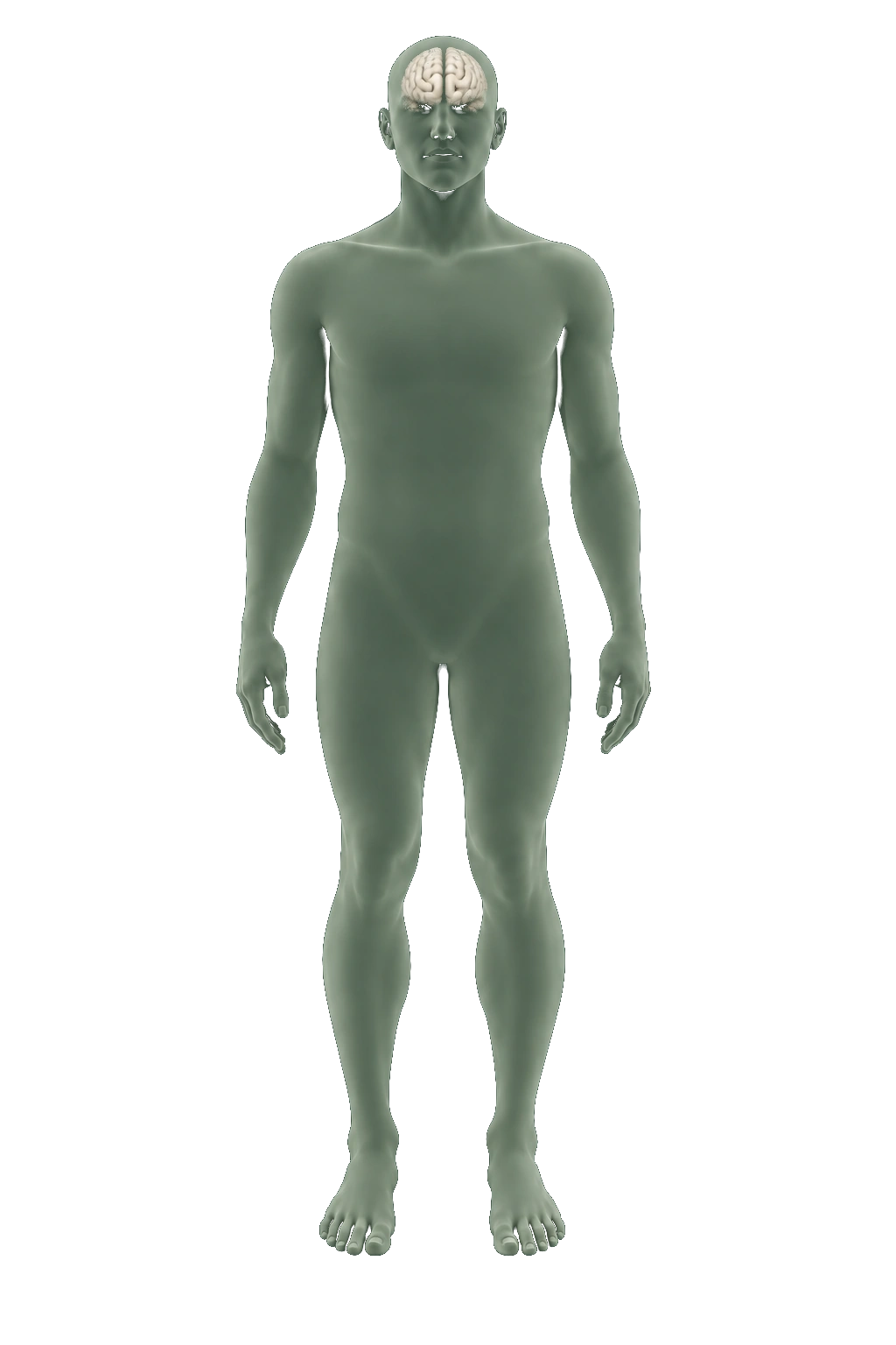
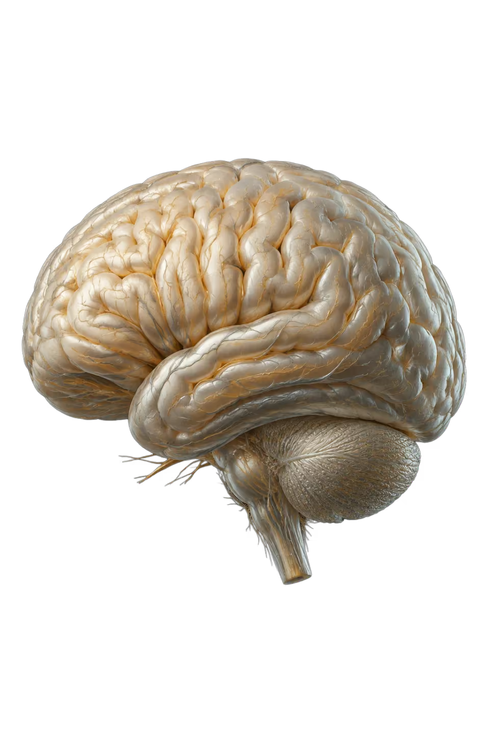
Brain anatomy

AI-generated educational illustration: Brain anatomy
About this anatomy

An isolated view of the brain. Select Lumbar spine to explore spinal imaging.

Choose another area ↑

Brain anatomy

Magnetic fields and radio waves create detailed internal images.

MRI does not use x-rays. Movement and some metal implants can affect the images.

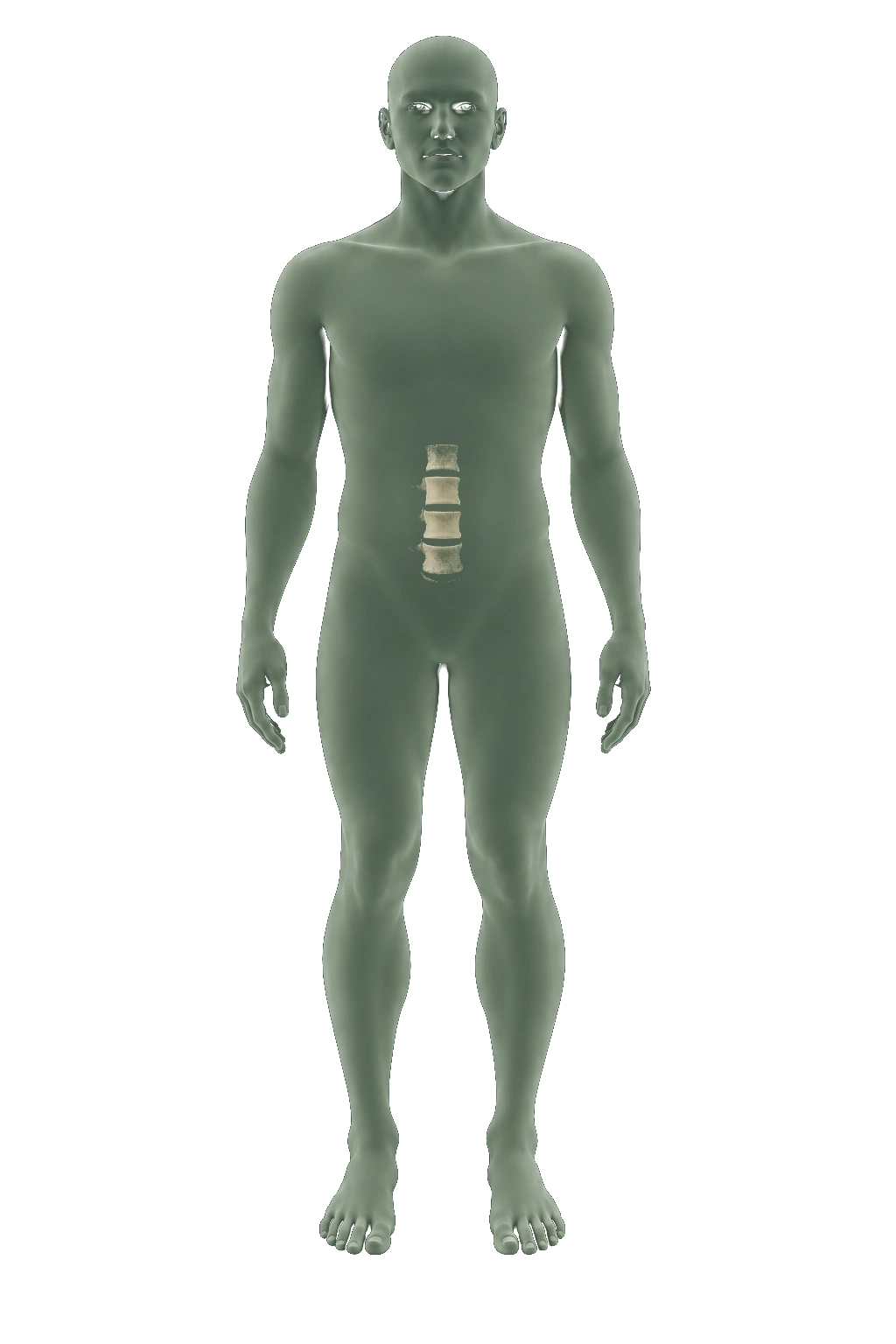
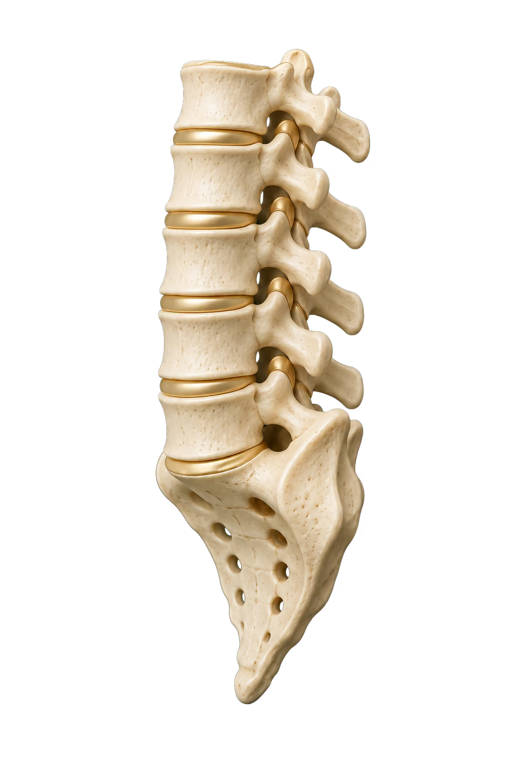
Lumbar spine

AI-generated educational illustration: Lumbar spine
About this anatomy

The lumbar vertebrae form the lower back, above the sacrum.

Choose another area ↑

Lumbar spine

Dedicated spine MRI shows discs, the spinal cord and surrounding soft tissues.

It can assess disc herniation and nerve compression. This illustration shows the lower spine; it is not an MRI image.

General MRI capability. A dedicated spine examination is not automatically included in whole-body screening; confirm EverVita’s protocol.

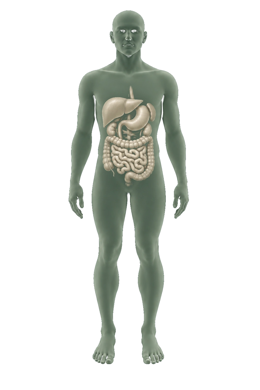
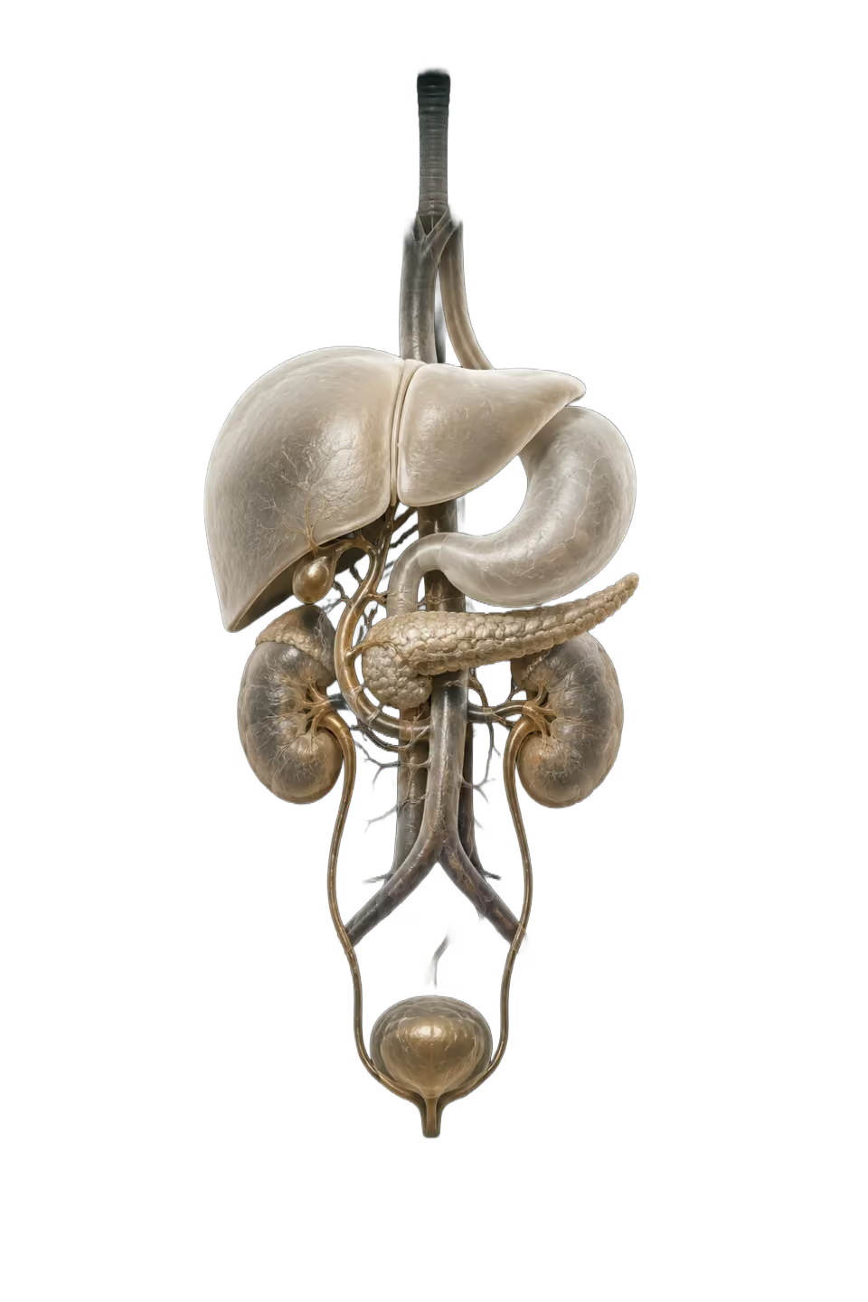
Abdominal anatomy

AI-generated educational illustration: Abdominal anatomy
About this anatomy

An overview of abdominal organs. Choose an individual organ for a closer view.

Choose another area ↑

Abdominal anatomy

Body MRI examines abdominal organs and pelvic structures.

It produces tissue images rather than bone-density measurements. Breathing and movement can blur abdominal images.

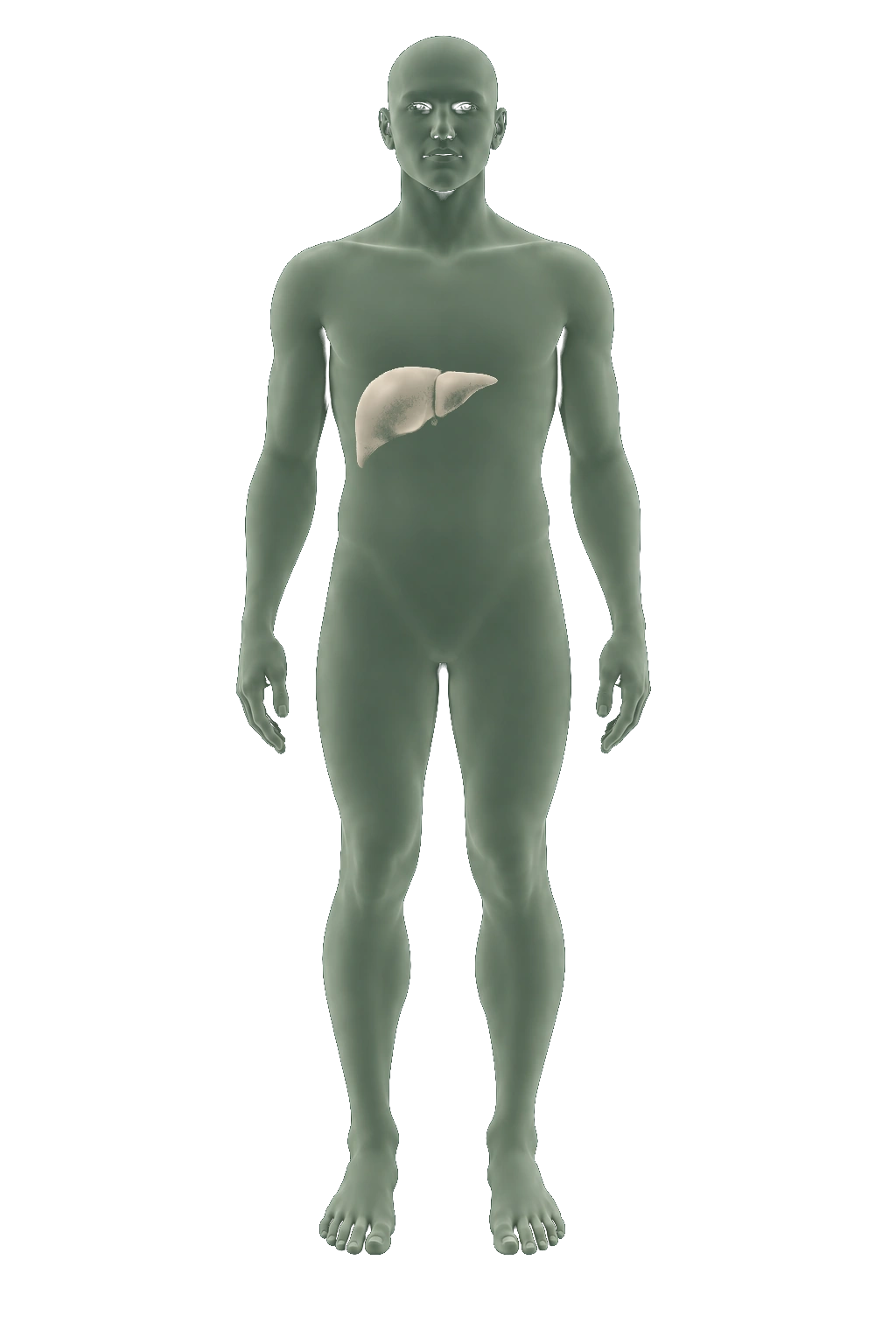
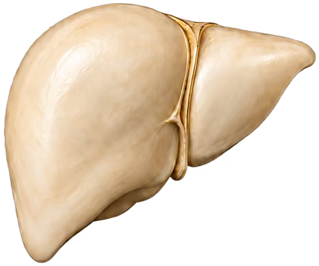
Liver

AI-generated educational illustration: Liver
About this anatomy

The liver makes bile to help digest fats and processes nutrients absorbed from food.

Choose another area ↑

Liver

Body MRI provides detailed soft-tissue views of the liver.

A radiologist reviews tissue detail and describes findings in a report. Additional views or a targeted examination may be needed.

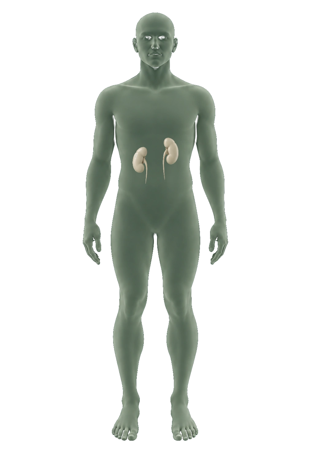
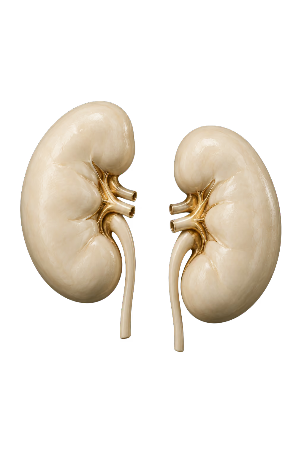
Kidneys

AI-generated educational illustration: Kidneys
About this anatomy

These paired organs filter blood, removing waste and excess water to make urine.

Choose another area ↑

Kidneys

Body MRI can examine the kidneys and surrounding structures.

Magnetic fields and radio waves create the images. A finding may need additional views or another imaging technique.

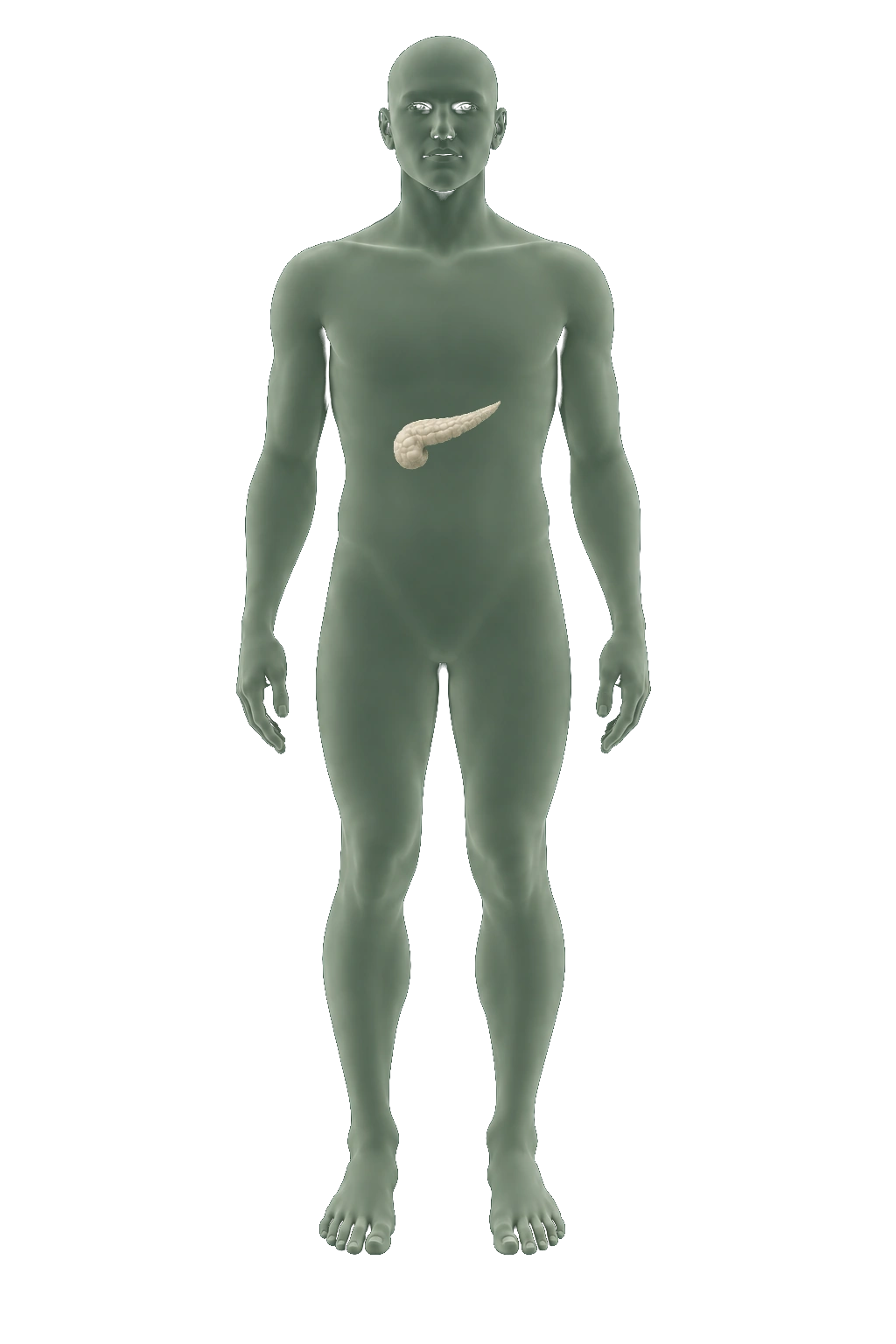
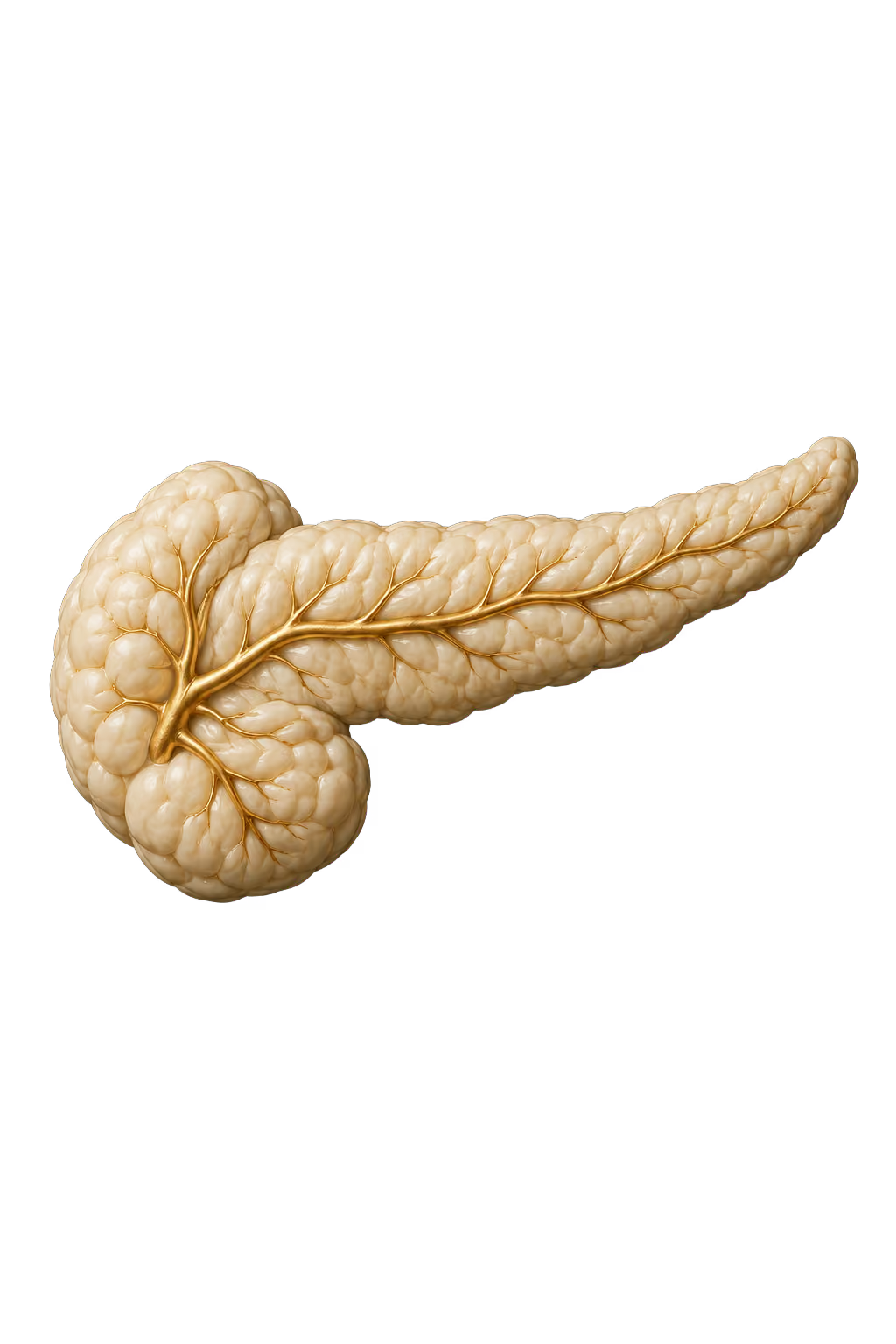
Pancreas

AI-generated educational illustration: Pancreas
About this anatomy

The pancreas produces digestive enzymes that help break down carbohydrates, fats and proteins.

Choose another area ↑

Pancreas

Body MRI is used to examine the pancreas.

MRI provides soft-tissue images without using x-rays. A targeted examination may be needed to investigate a finding.

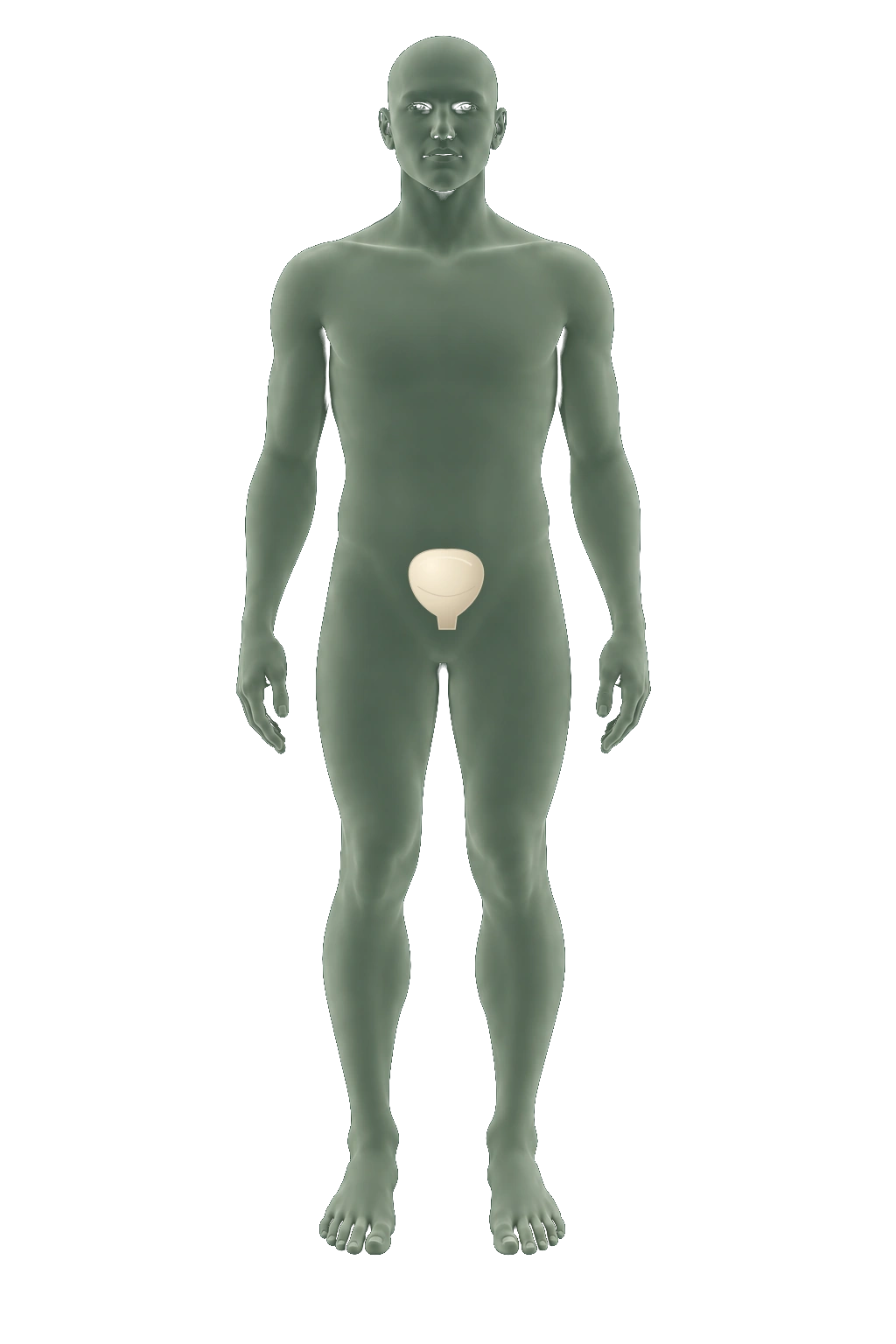
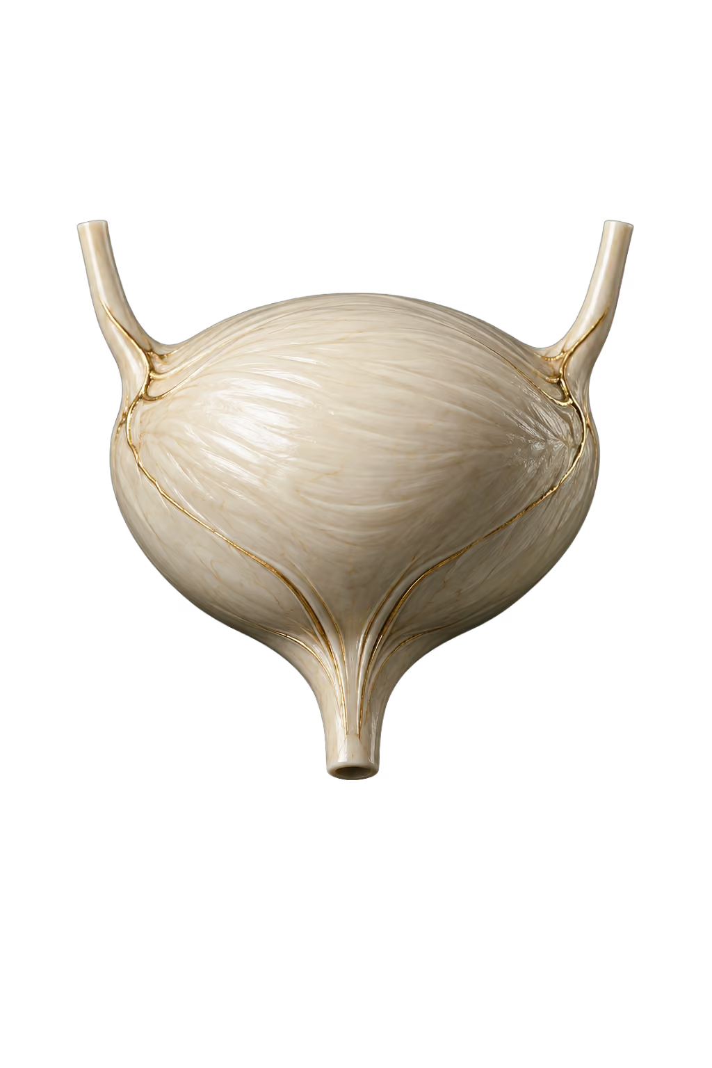
Bladder

AI-generated educational illustration: Bladder
About this anatomy

The bladder holds urine until it leaves the body.

Choose another area ↑

Bladder

Pelvic MRI can examine the bladder and nearby structures.

MRI creates soft-tissue images using magnetic fields and radio waves.

Coverage of this organ has not been confirmed. Check the package and protocol with the team.

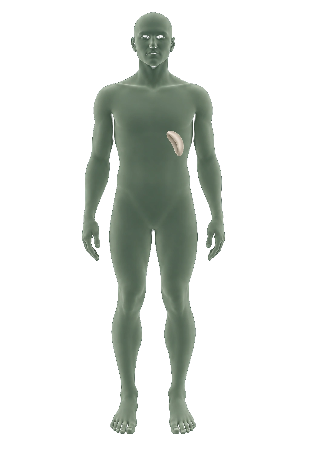
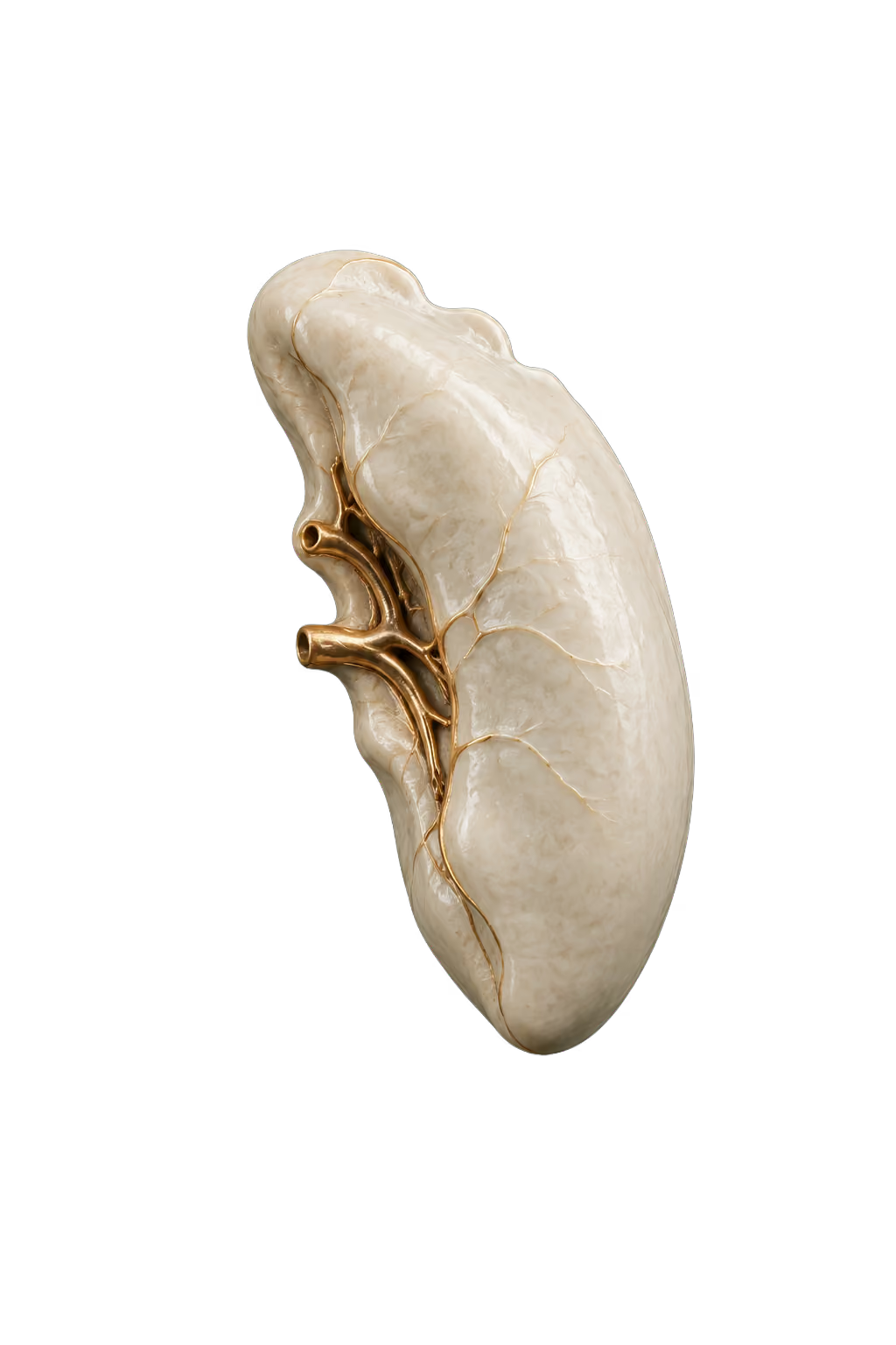
Spleen

AI-generated educational illustration: Spleen
About this anatomy

The spleen filters blood and removes old or damaged red blood cells.

Choose another area ↑

Spleen

Abdominal MRI can evaluate the spleen.

A radiologist interprets the images in the context of the examination.

Coverage of this organ has not been confirmed. Check the package and protocol with the team.

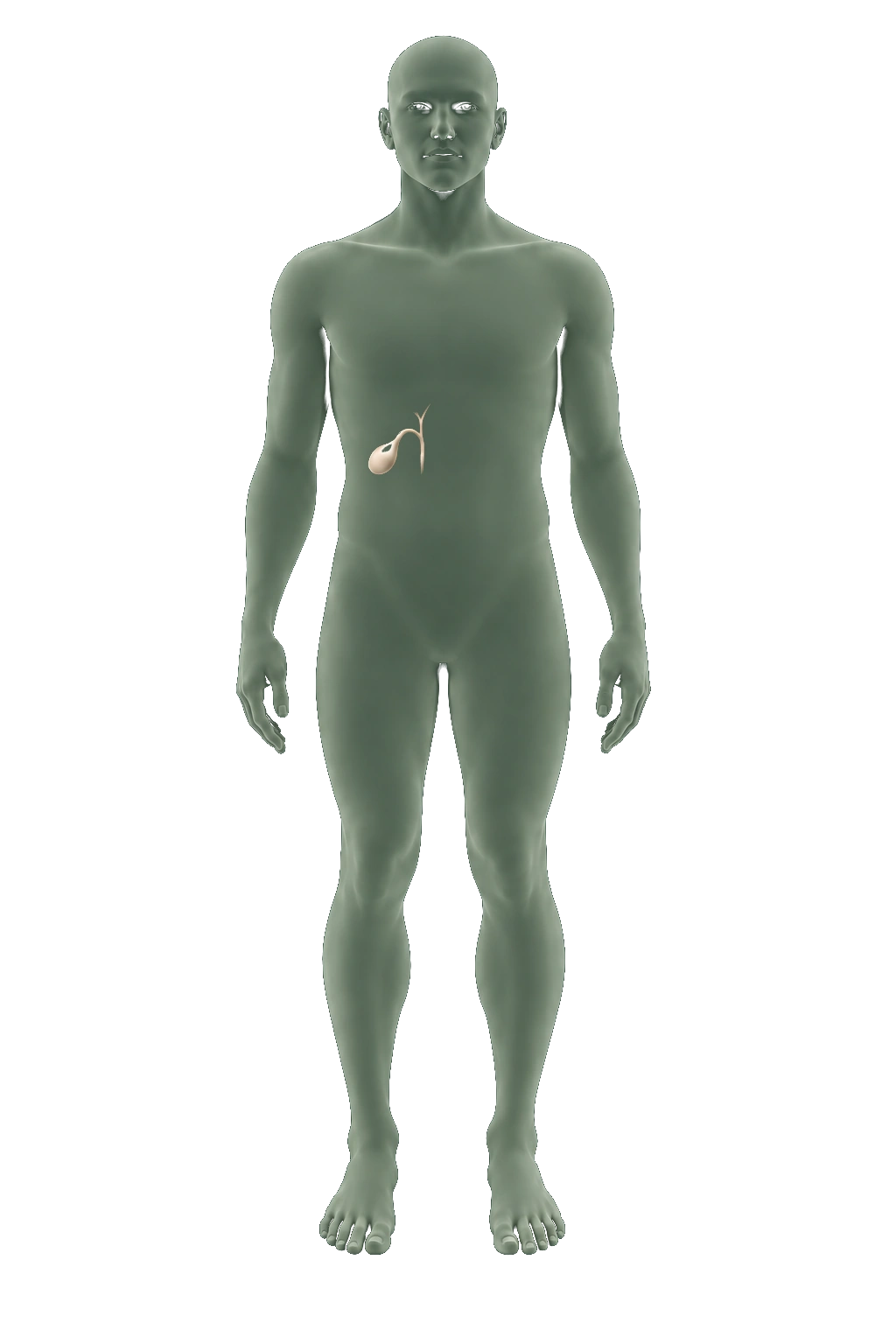
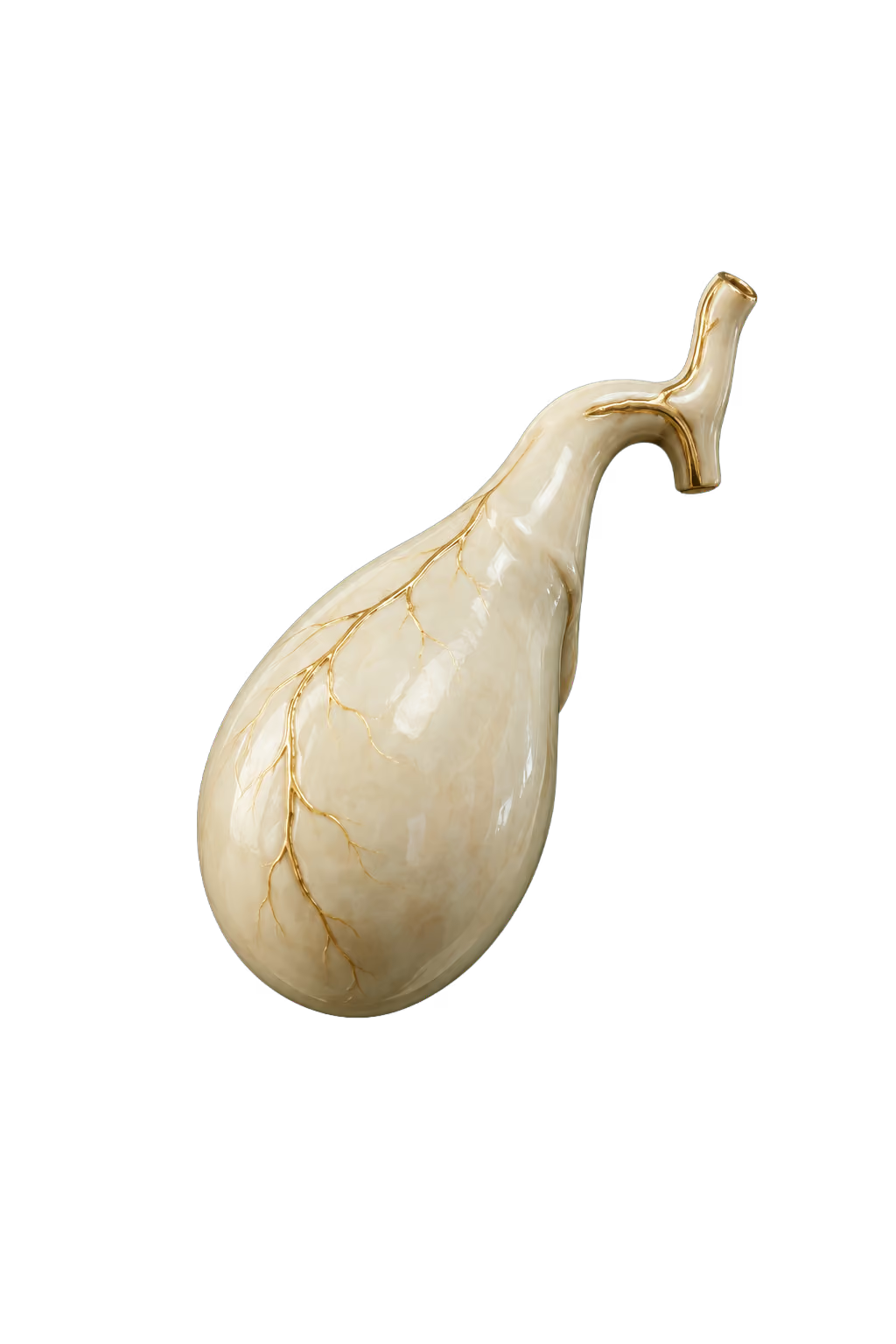
Gallbladder

AI-generated educational illustration: Gallbladder
About this anatomy

The gallbladder stores bile and releases it into the small intestine during digestion.

Choose another area ↑

Gallbladder

MRCP is a specialized MRI examination of the gallbladder and bile ducts.

A dedicated MRCP examination differs from a general whole-body MRI.

Coverage of this organ and this specialized examination has not been confirmed. Check the package and protocol with the team.

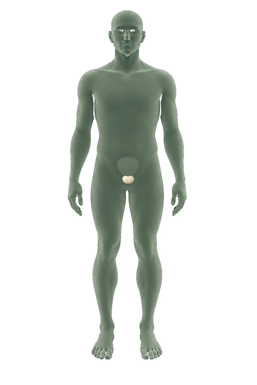
Prostate

AI-generated educational illustration: Prostate
About this anatomy

The prostate is a gland that contributes fluid to semen.

Choose another area ↑

Prostate

Dedicated prostate MRI provides detailed views of the gland.

Multiparametric MRI examines tissue structure, water movement and blood flow.

Coverage of this organ and this specialized examination has not been confirmed. Check the package and protocol with the team.

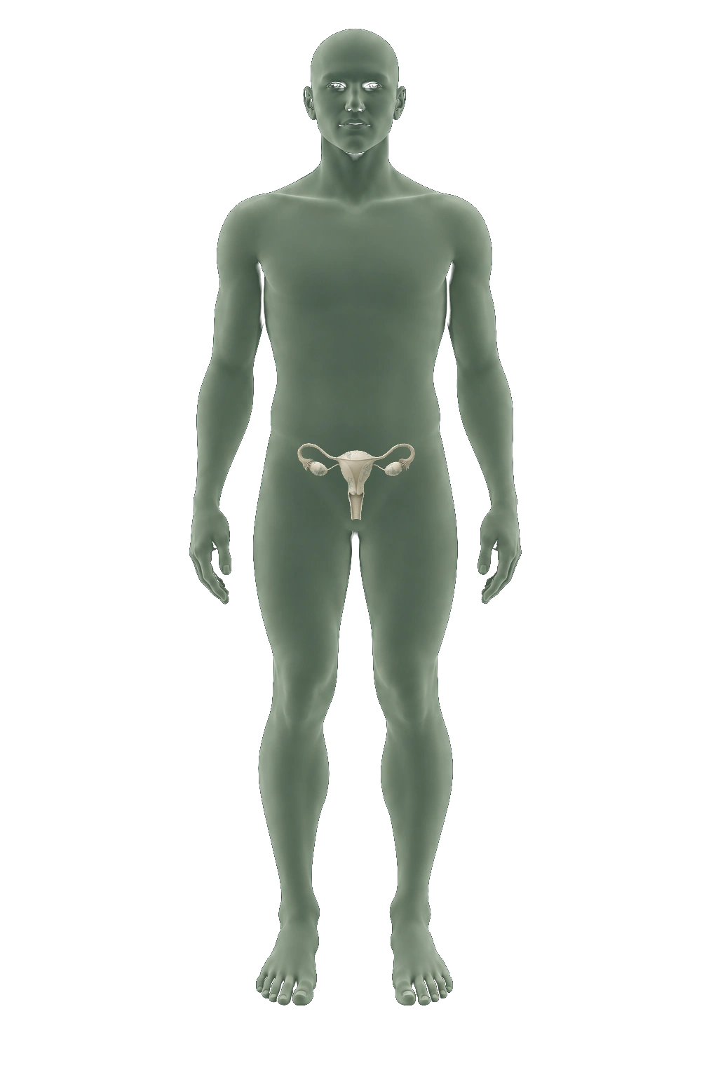
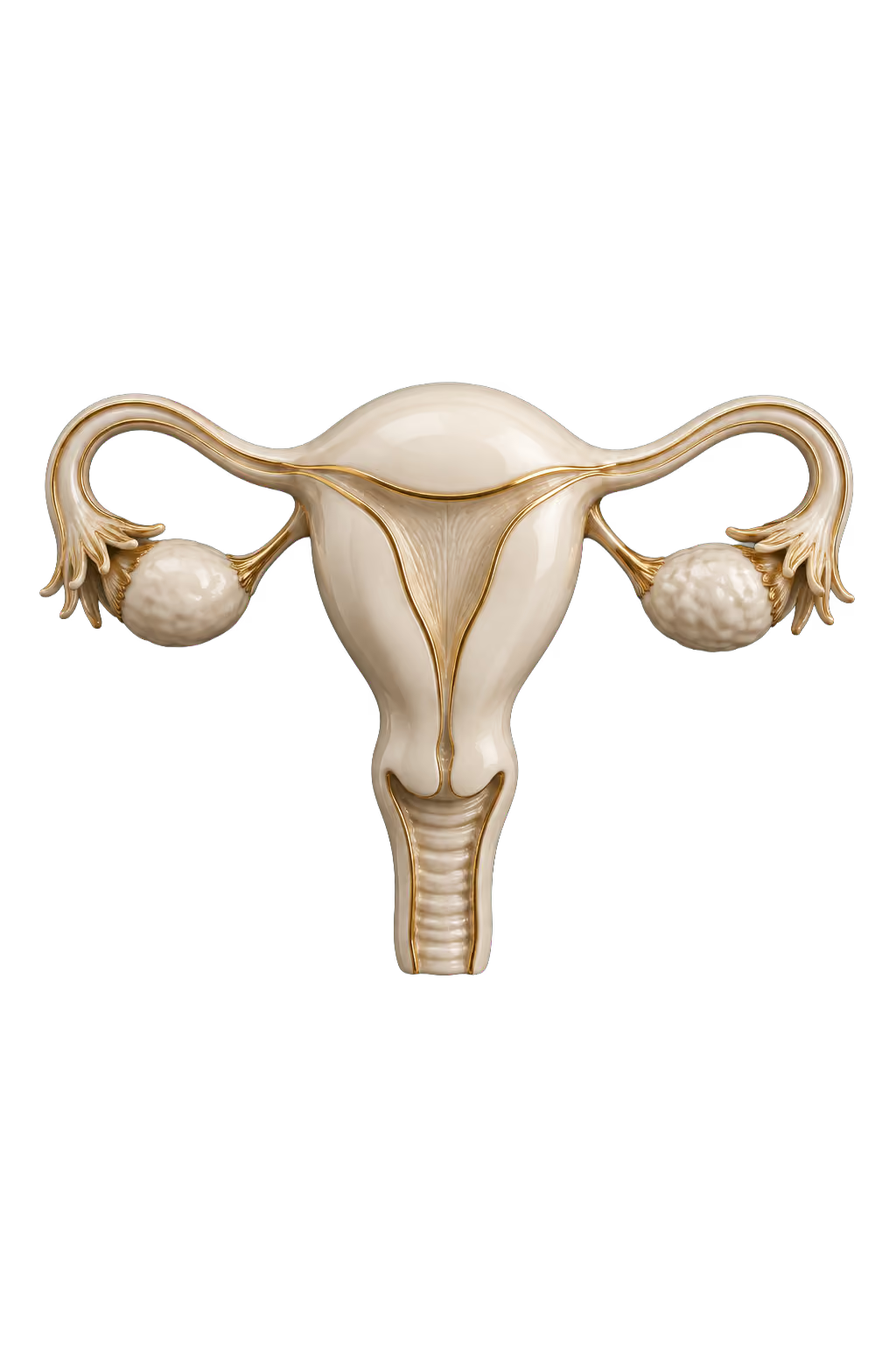
Uterus & ovaries

AI-generated educational illustration: Uterus & ovaries
About this anatomy

The uterus supports a developing pregnancy; the ovaries produce eggs and hormones.

Choose another area ↑

Uterus & ovaries

Pelvic MRI can evaluate the uterus and ovaries.

MRI provides soft-tissue detail without using x-rays.

Coverage of these organs has not been confirmed. Check the package and protocol with the team.

↑ Change scan

Organs & blood vessels

CT imaging

Explore what CT can show across the body. EverVita describes heart and lung examinations; the additional anatomy views explain general CT capabilities, not confirmed package inclusions.

Explore CT in detail
Illustrative body location, not a coverage map

Approximate location.
Not a coverage map.

Explore an area10 areas

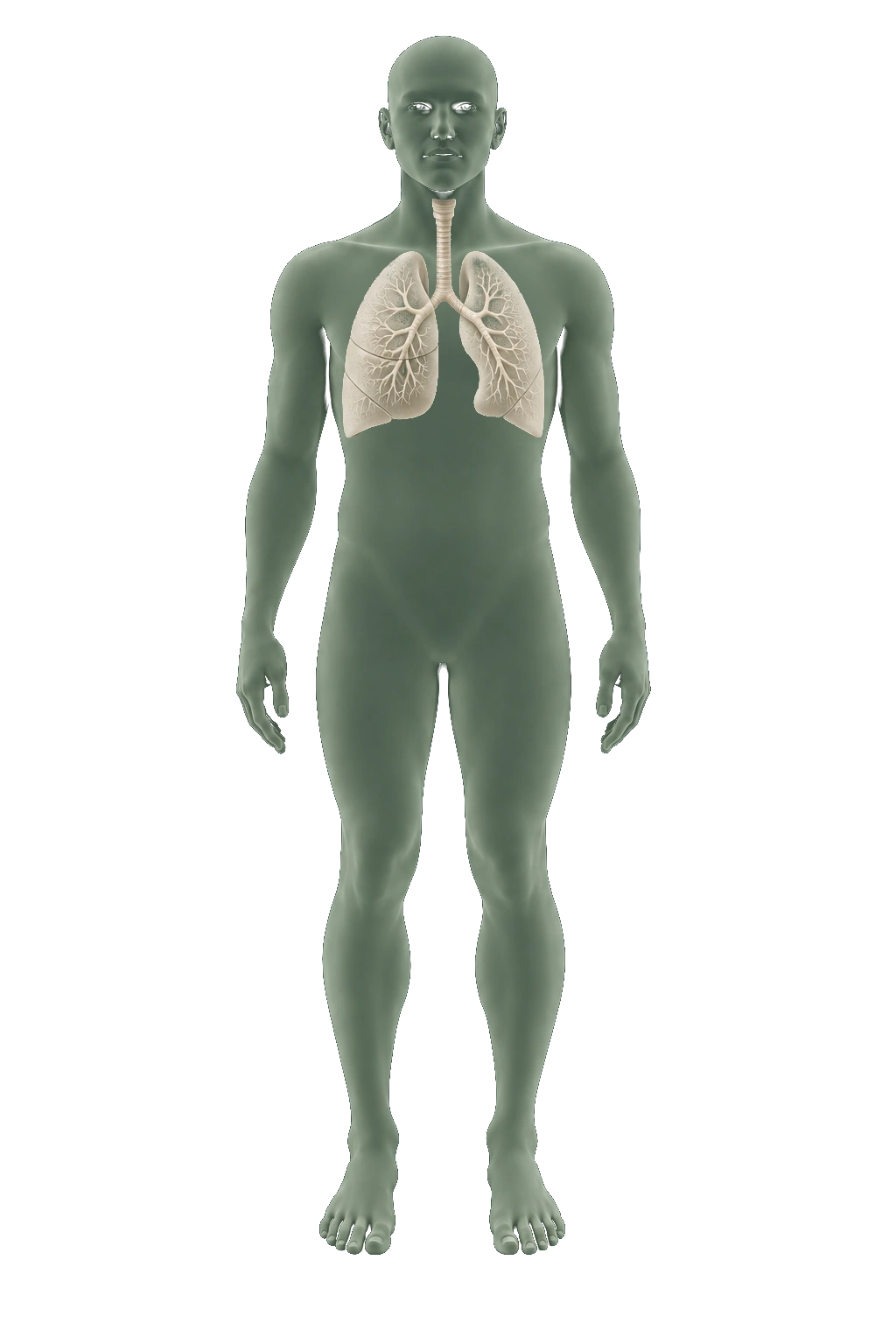
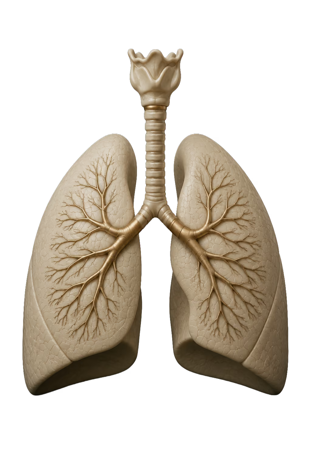
Lungs & airways

AI-generated educational illustration: Lungs & airways
About this anatomy

The paired lungs and branching airways are shown as an educational model.

Choose another area ↑

Lungs & airways

Cross-sectional chest images can reveal lung nodules.

Screening low-dose CT is performed without injected contrast. A nodule is not automatically cancer; further assessment may be needed.

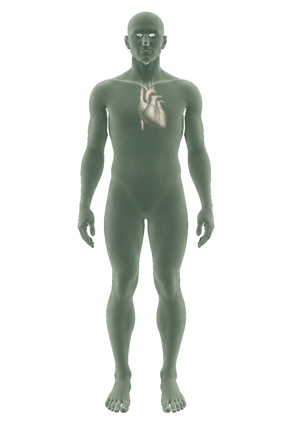
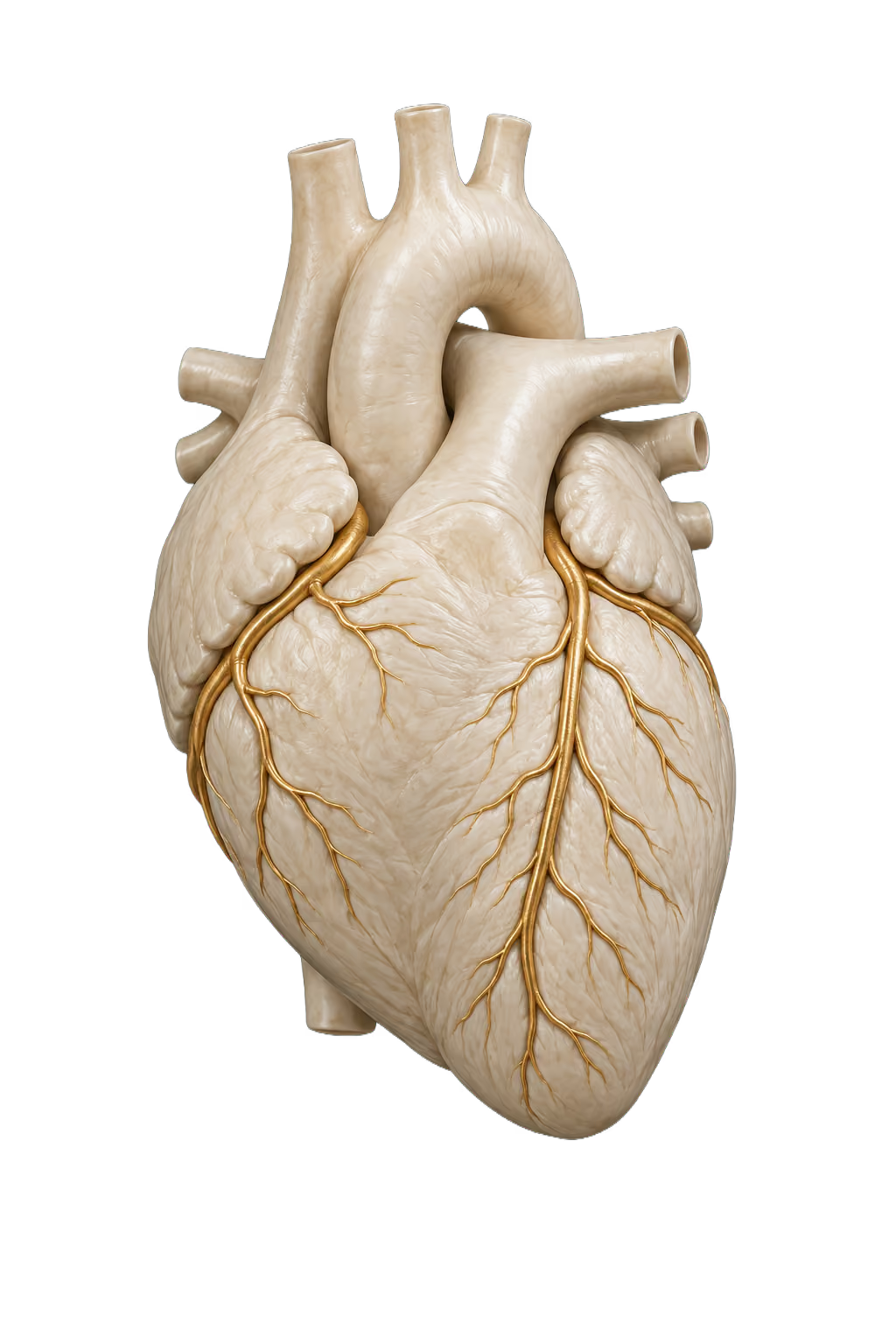
Heart

AI-generated educational illustration: Coronary arteries
About this anatomy

The coronary arteries run across the heart’s surface and supply its muscle.

Choose another area ↑

Heart & coronary arteries

Coronary CT angiography uses an injected iodine-containing contrast agent to examine the coronary arteries for plaque and narrowing.

The images help a clinician assess coronary artery disease. This is a diagnostic examination, not a treatment for a blockage.

Brain anatomy

AI-generated educational illustration: Brain anatomy
About this anatomy

An educational view of the brain.

Choose another area ↑

Brain anatomy

Head CT can reveal bleeding and injuries within the brain.

MRI can provide greater detail for some brain soft-tissue questions.

General CT capability. This area is not confirmed as part of EverVita’s heart or lung examinations.

Liver

AI-generated educational illustration: Liver
About this anatomy

The liver is shown as an isolated anatomical model.

Choose another area ↑

Liver

CT provides cross-sectional views of the liver and can help locate a mass or assess injury.

The examination technique depends on the clinical question.

General CT capability. This area is not confirmed as part of EverVita’s heart or lung examinations.

Kidneys

AI-generated educational illustration: Kidneys
About this anatomy

The paired kidneys and their urinary connections.

Choose another area ↑

Kidneys

CT can examine kidney masses, stones and urinary-tract abnormalities.

The examination technique depends on the question being investigated.

General CT capability. This area is not confirmed as part of EverVita’s heart or lung examinations.

Pancreas

AI-generated educational illustration: Pancreas
About this anatomy

An isolated view of pancreatic anatomy.

Choose another area ↑

Pancreas

Abdominal CT can investigate pancreatic inflammation and help locate pancreatic tumors.

The scan protocol is selected for the question being investigated.

General CT capability. This area is not confirmed as part of EverVita’s heart or lung examinations.

Spleen

AI-generated educational illustration: Spleen
About this anatomy

An isolated view of the spleen; CT examines it within the wider abdomen.

Choose another area ↑

Spleen

CT shows the spleen in cross-section alongside nearby abdominal organs.

A radiologist interprets these views in the context of the examination; a finding may need additional images or another technique.

General CT capability. This area is not confirmed as part of EverVita’s heart or lung examinations.

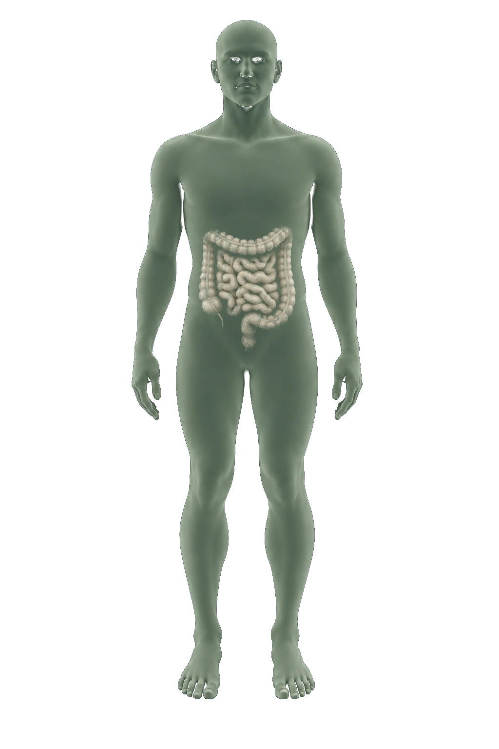
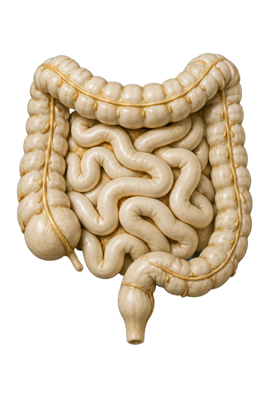
Bowel

AI-generated educational illustration: Bowel
About this anatomy

An illustrative view of the small and large intestines.

Choose another area ↑

Bowel

Abdominal CT can investigate bowel inflammation, including Crohn’s disease and diverticulitis.

Different bowel examinations use different preparation and imaging protocols.

General CT capability. This area is not confirmed as part of EverVita’s heart or lung examinations.

Bladder

AI-generated educational illustration: Bladder
About this anatomy

The bladder holds urine until it leaves the body.

Choose another area ↑

Bladder

CT urography examines the urinary tract, including the bladder, for abnormalities such as stones or tumors.

CT urography is a dedicated examination, not a feature of every CT scan.

General CT capability. This area is not confirmed as part of EverVita’s heart or lung examinations.

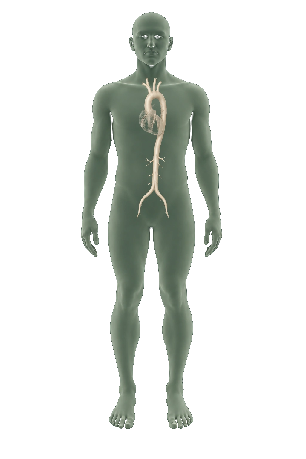
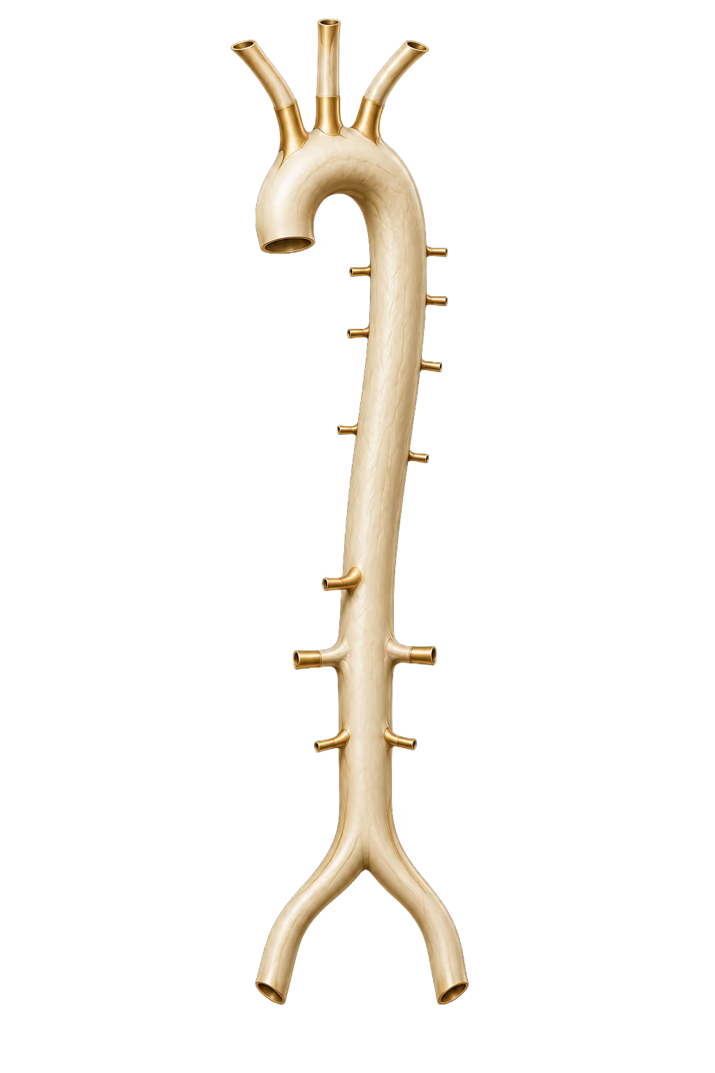
Aorta

AI-generated educational illustration: Aorta
About this anatomy

An illustrative view of the aortic arch and descending vessel.

Choose another area ↑

Aorta

CT angiography uses injected contrast to examine the aorta for aneurysms or tears in its wall.

The contrast timing and scan area are selected for the vessel being examined.

General CT capability. This area is not confirmed as part of EverVita’s heart or lung examinations.

↑ Change scan

Bone density & body composition

DXA

Explore bone-density sites and body-composition regions. EverVita describes both types of measurement; the exact sites and regional reporting available in your examination need confirmation.

Explore DXA in detail
Illustrative body location, not a coverage map

Approximate location.
Not a coverage map.

Explore an area6 areas

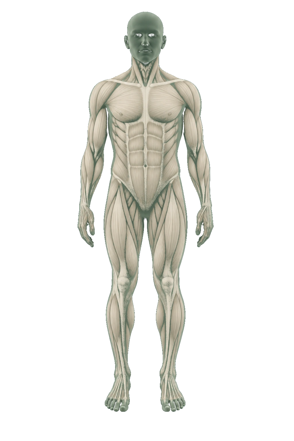
Body composition

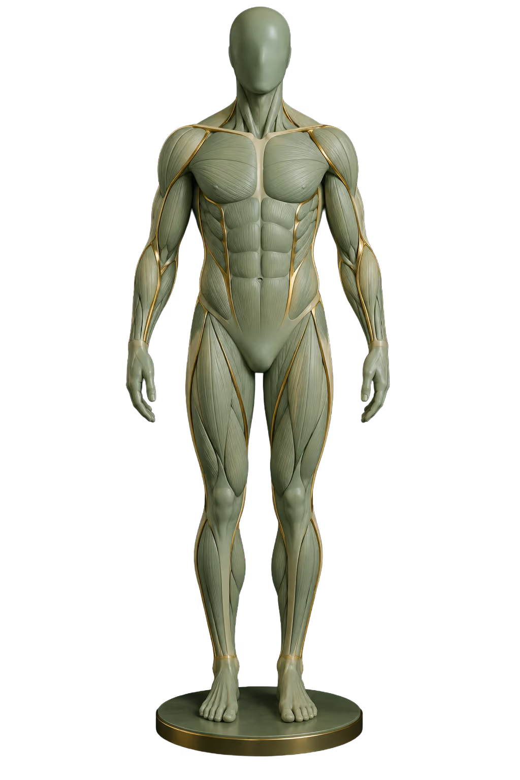
AI-generated educational illustration: Body composition
About this anatomy

A stylized soft-tissue model. DXA distinguishes fat and lean tissue; the illustration contains no personal measurements.

Choose another area ↑

Body composition

Fat mass, lean mass and body-fat percentage.

Separates tissue measurements that a scale combines into one number. Scanner and analysis differences matter when comparing repeat measurements.

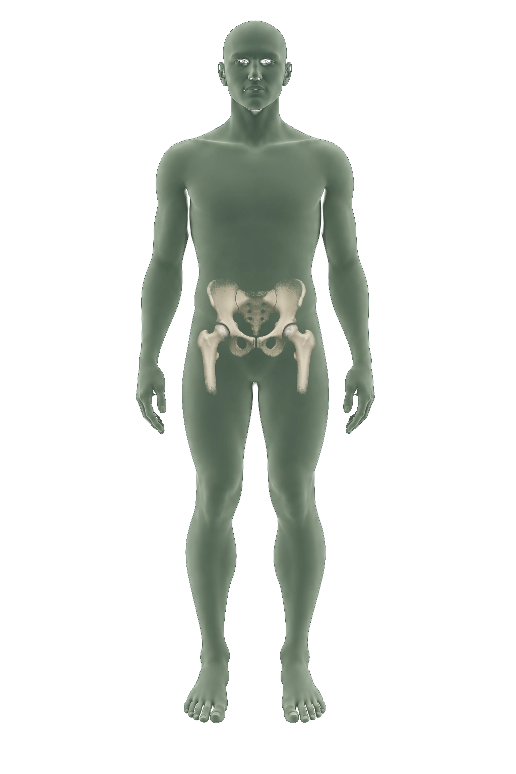
Hip

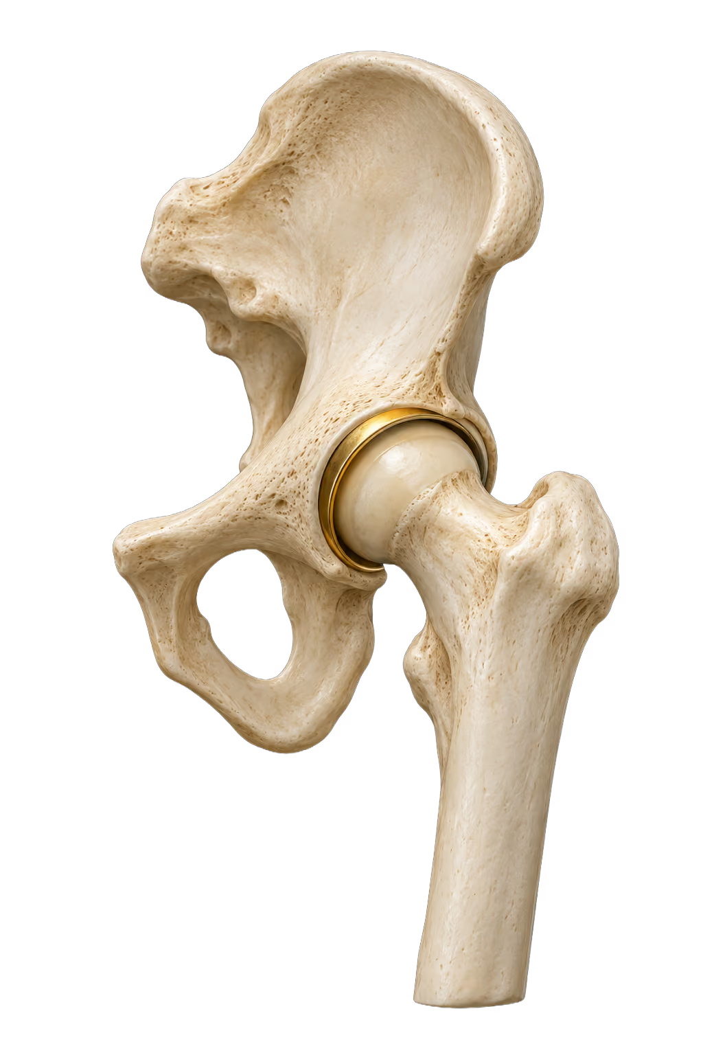
AI-generated educational illustration: Hip
About this anatomy

The hip joint connects the head of the thigh bone to a socket in the pelvis.

Choose another area ↑

Hip

The hip is a common site for measuring bone density. The forearm is an alternative site in selected clinical circumstances.

Measurements are compared with reference populations using T-scores or Z-scores. Density helps assess fracture risk; it cannot predict who will have a fracture.

Confirm which measurement sites are included in your EverVita examination.

Lumbar spine

AI-generated educational illustration: Lumbar spine
About this anatomy

The lumbar vertebrae form the lower back, above the sacrum.

Choose another area ↑

Lumbar spine

The lower spine is another common site for a DXA bone-density measurement.

Additional assessments

Trabecular bone score (TBS) adds information to fracture-risk assessment. A separate vertebral fracture assessment (VFA) looks for spinal compression fractures. These do not assess bulging discs.

TBS and VFA require additional analysis or imaging. Confirm availability and measurement sites with EverVita.

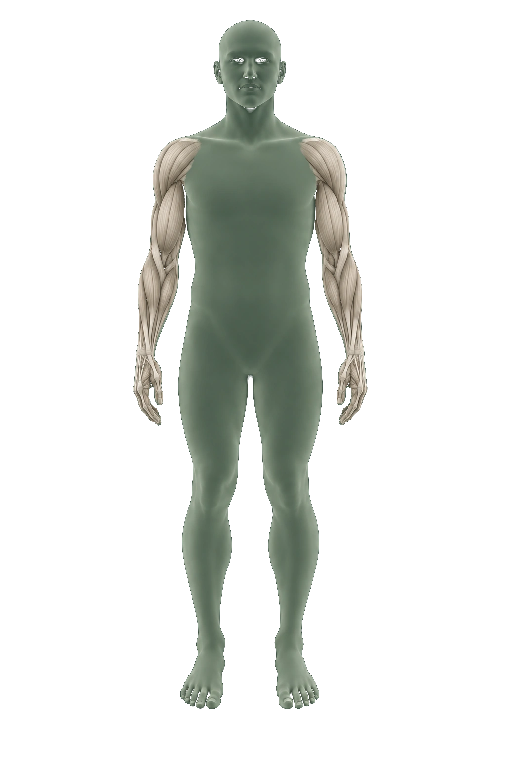
Arm composition

AI-generated educational illustration: Arm composition
About this anatomy

A soft-tissue view of the arm. Colors and proportions are illustrative, not measured results.

Choose another area ↑

Arm composition

Regional body-composition analysis can report fat and lean tissue for each arm.

Arm and leg lean-tissue measurements together contribute to assessing low muscle mass. They do not measure strength or diagnose sarcopenia on their own.

General DXA capability. Confirm whether this measurement is available in your EverVita examination.

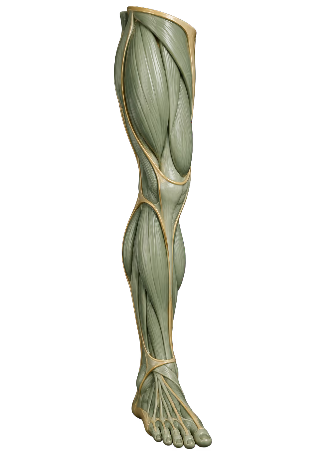
Leg composition

AI-generated educational illustration: Leg composition
About this anatomy

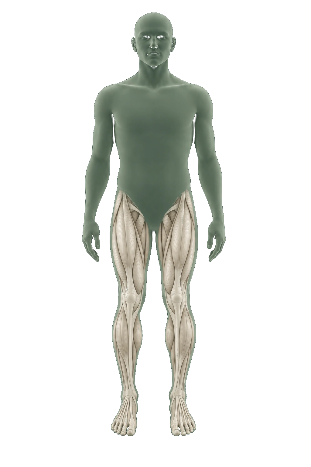
A soft-tissue view of the leg. Colors and proportions are illustrative, not measured results.

Choose another area ↑

Leg composition

Regional body-composition analysis can report fat and lean tissue for each leg.

These measurements describe lower-limb composition, not walking ability or muscle strength. Comparing changes requires consistent scanning and analysis.

General DXA capability. Confirm whether this measurement is available in your EverVita examination.

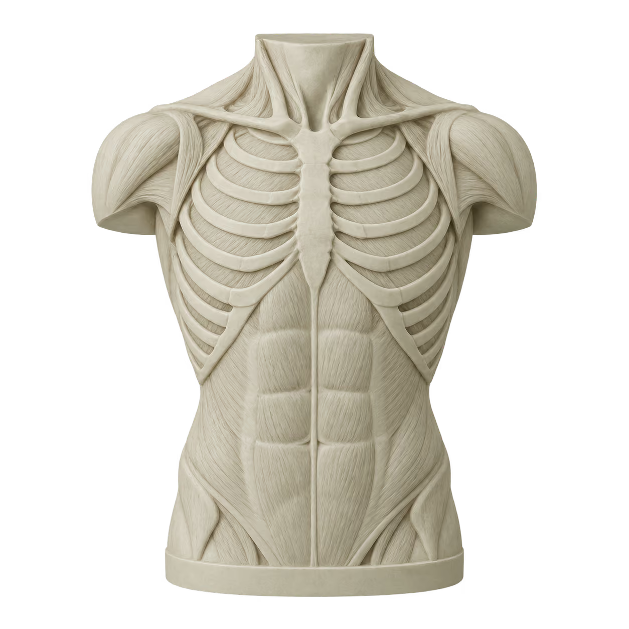
Abdominal composition

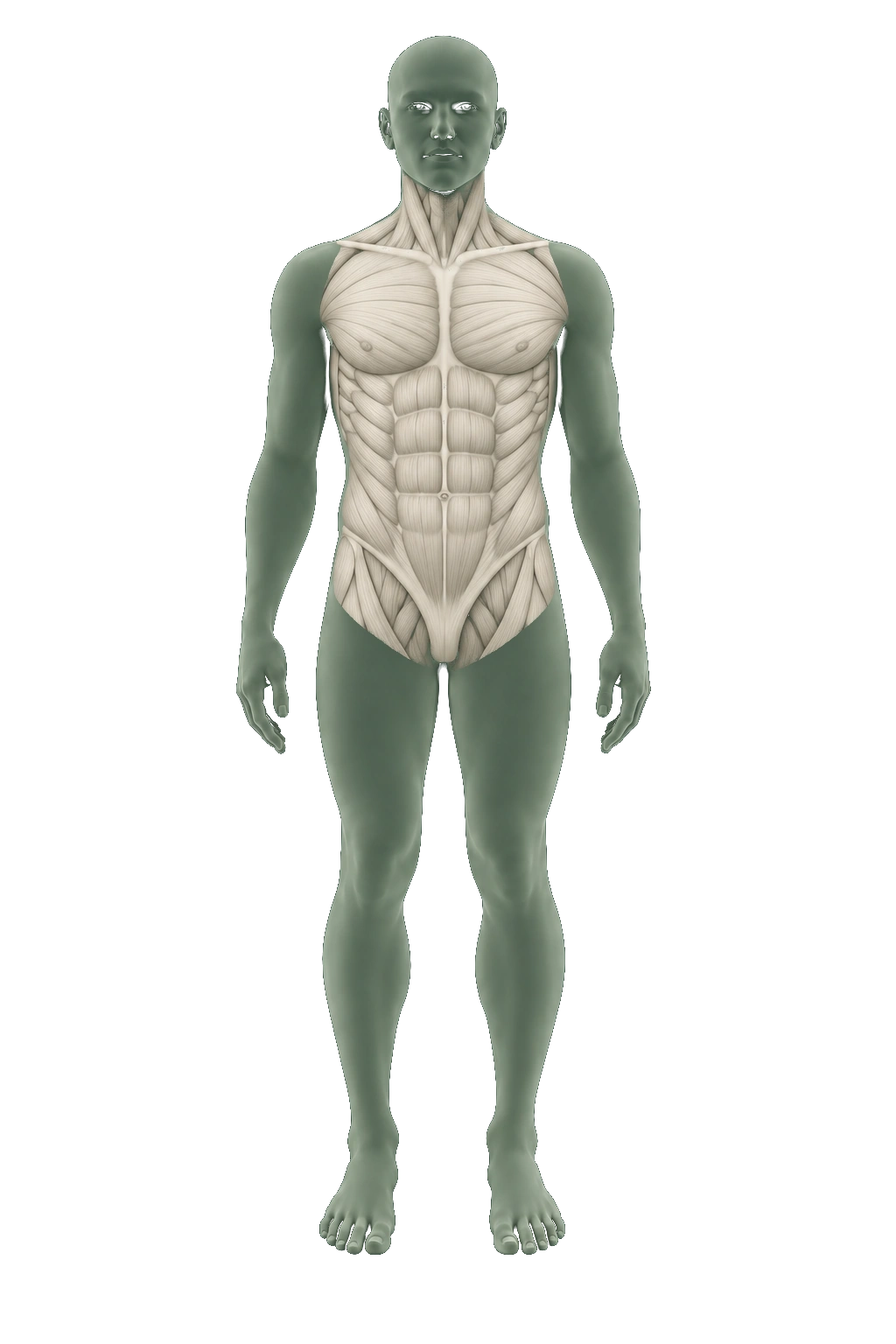
AI-generated educational illustration: Abdominal composition
About this anatomy

A torso-focused view of the soft-tissue model; not a measured fat distribution.

Choose another area ↑

Abdominal composition

Regional DXA analysis describes fat and lean tissue in the trunk, which includes the abdomen.

Compatible software can separately estimate visceral fat around abdominal organs. This is not the same as total trunk fat or an examination of organ health.

Visceral-fat estimation requires compatible analysis software. Confirm which regional measurements EverVita provides.

AI-generated anatomy illustrations · not diagnostic imaging. General examination information; confirm EverVita’s package coverage and protocol with the team.

Ready to compare what’s included?

Compare packages & pricing →
Questions for your appointment
  • How will my results be explained?
  • What consultation and follow-up are included in the package?
  • Who should I contact if I have questions afterward?
About this guide & sources

Service summaries reflect EverVita’s published descriptions, reviewed September 8, 2026. They do not confirm an individual package or clinical protocol. Illustrations are conceptual and not clinically validated.

EverVita MRI · EverVita CT · EverVita DXA

Scan explanations: Body MRI · CCTA · Lung screening · Bone density · DXA positions & additional analyses.

Organ explanations: NIDDK: kidneys · NIDDK: digestion · Urinary tract · Spleen · Prostate · Uterus and ovaries.

Targeted imaging: Spine MRI · Abdominal and pelvic MRI · MRCP · Prostate MRI. These references describe general examinations, not confirmed EverVita inclusions.

General capabilities: Head CT · Body CT · Abdominal CT · CT urography · CT angiography · CDC: regional DXA measurements. These describe general uses, not EverVita package coverage.

MRI / Brain selected.

A specific area. A focused perspective.

Focused scans.

Focused MRI and CT examinations concentrate on a particular area or imaging question. Compare availability and inclusions on the pricing page.

Focused preventive MRI

Single Body Part Scan

A focused preventive MRI of one body part. Confirm the region and preparation with the team.

View options & pricing

Additional assessments.

Eye, dental, blood and gut assessments offer information beyond the MRI, CT and DXA examinations.

Conceptual eye model

Retinal OCT

See the layers beneath the surface of your retina, with detailed cross-sectional images that help assess retinal health.

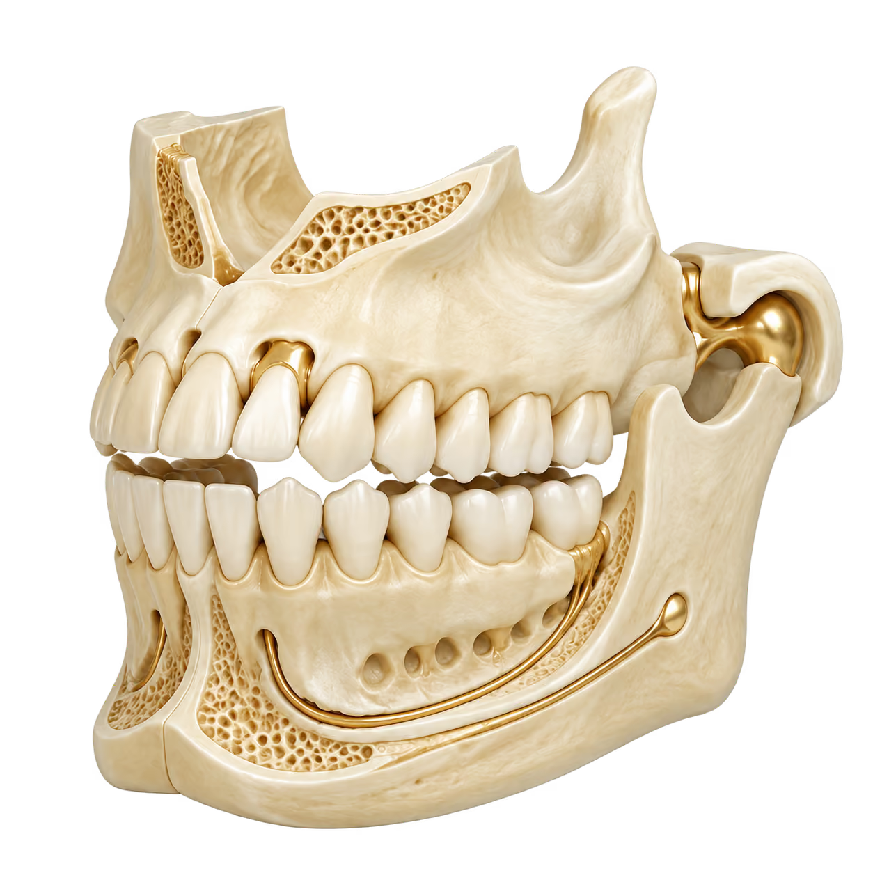
Conceptual teeth and jaw model

Dental CBCT

Look beneath the surface with three-dimensional images of your teeth, roots and jawbone.

Conceptual laboratory sample tubes

Blood biomarkers

Go beyond anatomy with blood-based measurements that add context to your health assessment.

Conceptual intestinal anatomy

Gut health testing

Explore the microbes in your gut with a stool-based microbiome test from Tiny Health.

These assessments are not included in every package. Confirm which are available in your chosen assessment and how to prepare.

Conceptual illustrations

Assessment questions →
Illustrative conversation in a warm setting
Your experience

Your visit,
thoughtfully arranged.

Before you arrive, during your appointment and when you review your results.

Before your visit

Follow the preparation instructions for your booked examinations. Ask the team about medication, fasting or implant-related questions before your visit.

At your appointment

Confirm which examinations are scheduled and ask what each involves before it begins.

Reviewing your results

Discuss the findings with your reviewing physician and ask whether additional testing or follow-up is recommended.

Your results

EverVita’s main packages list radiologist interpretation and a physician consultation. Compare packages for the examinations and review arrangements included.

View packages & pricing

Confirm when and how you will receive your report and whether follow-up visits are included. Imagery is illustrative.

Preparation & results FAQs →

Before you book

Choose with
a clearer picture.

Your package determines the examinations and assessments included. Confirm the inclusions, preparation and results review before booking.

Discuss your options
How do I choose a package?

Confirm which MRI, CT or DXA examinations and additional assessments are included in the package you are considering. Discuss suitability with the team; the anatomy explorer is a general guide, not a package coverage map.

How should I prepare?

Follow the instructions provided for your chosen appointment. Confirm any preparation questions with the team before your visit.

What happens after my scan?

Confirm how you will receive your report, how your results will be reviewed, and which consultation or follow-up arrangements are included in your package.

Is screening suitable for me?

Discuss suitability with a qualified clinician. Screening does not replace evaluation of symptoms or routine recommended care.

Your scans. Your next step.

Compare package inclusions and pricing, or contact the team for help choosing.

Compare packages & pricing Not sure where to start? Talk to our team →

Educational illustration

AI-generated anatomy. Not a diagnostic scan or patient result.