




The findings guide
What yourpreventive healthassessmentmay show
Explore plain-language explanations of imaging findings and selected health measurements. Understand what a term may mean—and why your personal context matters.
Explore the library
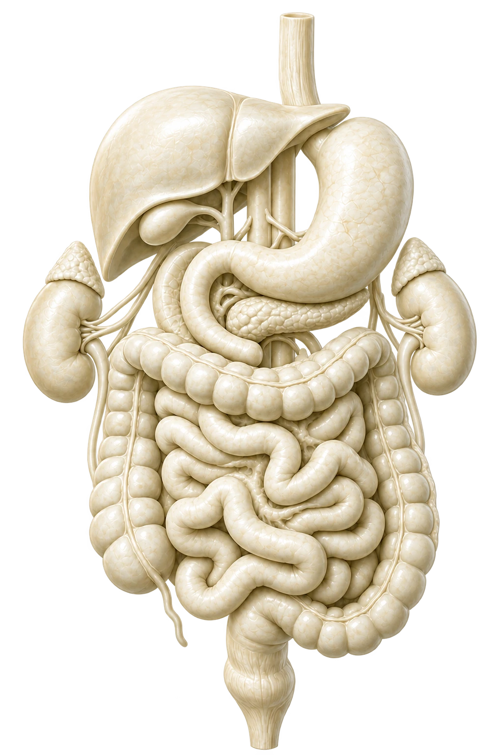
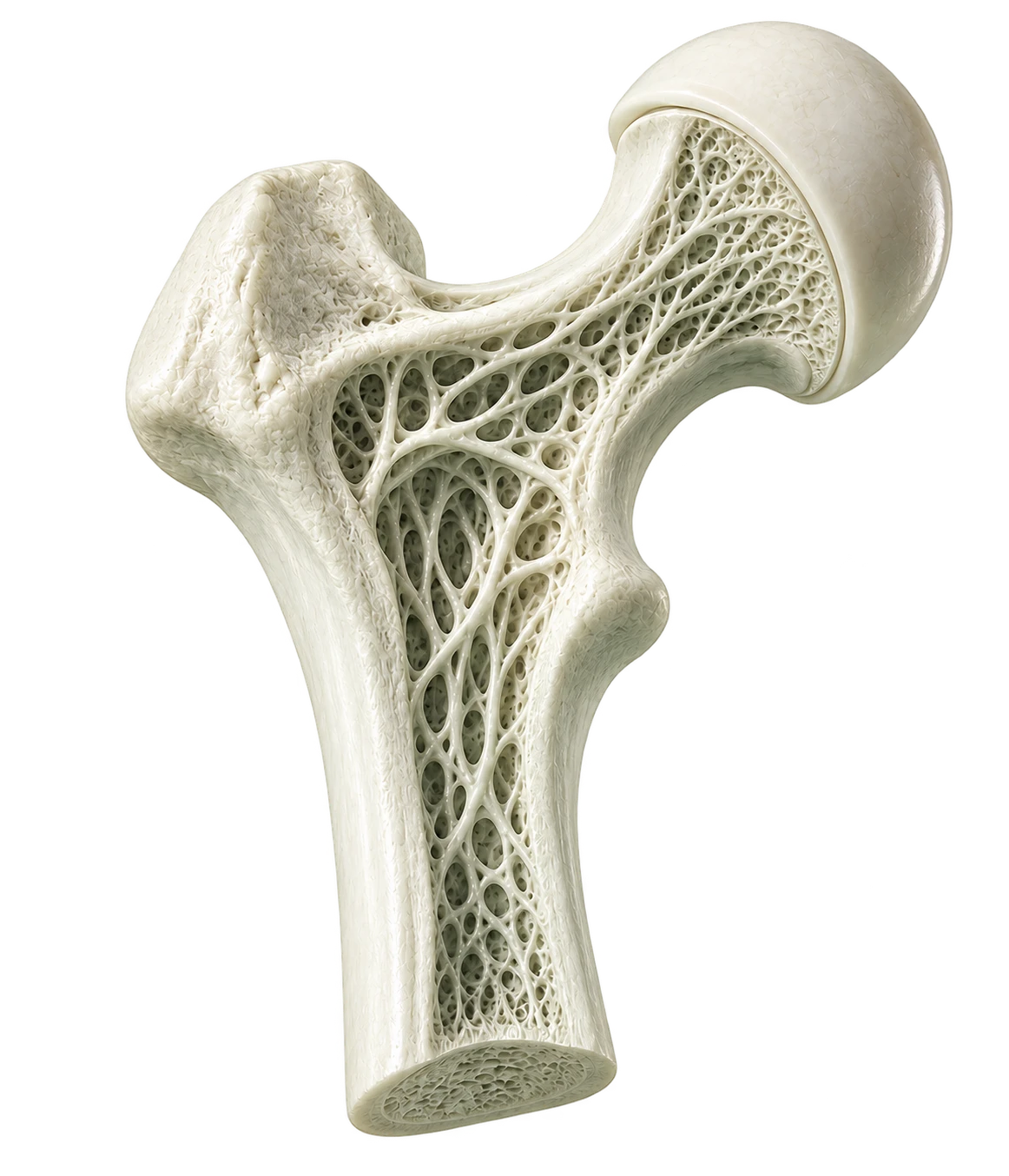

Explore with clarity
Explore the findings library
Brain & Neurologic Health
Acoustic Schwannoma
Brain
An acoustic schwannoma is usually a noncancerous tumor that develops along a nerve involved in hearing and balance. MRI may identify the growth and show its relationship to nearby structures.
Arachnoid Cyst
Brain
An arachnoid cyst is a fluid-filled sac that forms within one of the coverings around the brain or spinal cord. Many are harmless and found incidentally.
Arteriovenous Malformation (AVM)
Brain
An arteriovenous malformation is an unusual connection between arteries and veins. Imaging can show the abnormal blood vessels and their relationship to nearby brain tissue.
Brain Abscess
Brain
A brain abscess is a collection of infected material within the brain. Imaging may identify the location and extent of the abnormality, while urgent clinical evaluation is needed to determine the cause and treatment.
Brain Aneurysm
Brain
A brain aneurysm is a weakened area in a brain artery that has widened or bulged. Imaging can show its location and size, while follow-up depends on the aneurysm's appearance and clinical context.
Brain Atrophy or Volume Loss
Brain
A reduction in brain tissue volume that may be related to aging or another neurologic condition.
Brain Bleed (Hemorrhage)
Brain
A brain hemorrhage is bleeding within or around the brain. Imaging can help identify the location and extent of bleeding and whether urgent evaluation is needed.
Brain Cyst
Brain
A fluid-filled area within or near the brain that is often harmless but may need review depending on its location and size.
Brain Mass or Tumor
Brain
A brain mass or tumor is an area of abnormal tissue within or near the brain. Imaging can show its location and effects on nearby structures, while additional testing may be needed to determine its type.
Brain Metastases
Brain
Brain metastases are cancer deposits that have spread to the brain from another part of the body. Imaging may identify one or more brain lesions, but additional evaluation is needed to determine their cause and guide care.
Brain Volume Assessment
Brain
A brain volume assessment estimates the size of selected brain regions and may compare results with expected ranges for age or other reference groups. It is a health metric, not a diagnosis by itself, and should be interpreted with clinical context.
Carotid Artery Narrowing (Stenosis)
Head, Neck & Throat
Carotid artery narrowing, also called stenosis, is a reduction in the inner space of an artery supplying the brain. Neck vascular imaging may identify plaque or narrowing, while clinical evaluation determines its significance and next steps.
Chiari Malformation
Brain
A structural difference where part of the lower brain extends farther toward the spinal canal than usual.
Enlarged Brain Ventricles
Brain
Widening of the fluid-filled spaces within the brain, which may or may not reflect excess fluid pressure.
Glioma
Brain
A glioma is a tumor that develops from supporting cells in the brain or spinal cord. Imaging may identify a suspicious mass, while additional testing is needed to determine the exact type and grade.
Hydrocephalus
Brain
Hydrocephalus is an abnormal buildup of fluid within the spaces of the brain. It can enlarge the brain’s ventricles and may require medical evaluation depending on symptoms and severity.
Meningioma
Brain
A meningioma is usually a slow-growing tumor that develops from the tissues covering the brain and spinal cord. Many are noncancerous and discovered incidentally on imaging.
Multiple Sclerosis (MS)
Brain
Multiple sclerosis is a condition in which the immune system damages the protective covering of nerves in the brain and spinal cord. MRI may show characteristic areas of demyelination, although diagnosis requires clinical evaluation.
Neurocysticercosis
Brain
Neurocysticercosis is an infection that can form small cyst-like lesions in the brain. Imaging may show characteristic lesions or calcifications that require clinical evaluation.
Pineal Cyst
Brain
A pineal cyst is a fluid-filled area in the pineal gland near the center of the brain. Most are small, harmless, and found incidentally.
Pituitary Adenoma
Brain
A pituitary adenoma is a usually noncancerous growth of the pituitary gland. Imaging may show the growth and its relationship to nearby structures, while hormone testing may be used for further evaluation.
Pituitary Mass
Brain
A pituitary mass is an abnormal growth involving the pituitary gland. Many are noncancerous, but size, hormone effects, and imaging appearance determine whether further evaluation is needed.
Small Vessel Ischemic Changes
Brain
Small vessel ischemic changes affect the brain's smaller blood vessels. They are commonly seen with aging and vascular risk factors and may appear as changes in the brain's white matter.
Spinal Cord Infarct
Spine
A spinal cord infarct is an area of spinal cord injury caused by reduced blood flow. MRI may show the location and extent of the injury.
Spinal Cord Lesion
Spine
An area of abnormal tissue or signal involving the spinal cord that may need further evaluation.
Split Cord Malformation
Spine
Split cord malformation is a congenital condition in which part of the spinal cord is divided into two sections. MRI can show the anatomy of the spinal cord and surrounding structures.
Stroke (Brain Infarct)
Brain
A brain infarct is an area of tissue injury caused by reduced blood flow, as occurs with an ischemic stroke. Imaging may show recent or older areas of injury.
Syrinx (Syringomyelia)
Spine
A syrinx is a fluid-filled cavity within the spinal cord. MRI can show its location and extent and whether it is associated with other spinal or brain abnormalities.
Tarlov Cyst
Spine
A Tarlov cyst is a fluid-filled sac that forms around a nerve root, usually in the lower spine. Many are found incidentally and cause no symptoms.
Tethered Cord
Spine
A spinal cord that is attached more tightly than usual and may not move freely within the spinal canal.
Transverse Myelitis
Spine
Transverse myelitis is inflammation involving part of the spinal cord. MRI may show areas of abnormal signal within the cord, while clinical evaluation and additional testing are needed to determine the cause.
Heart & Vascular Health
Aortic Aneurysm
Heart & Blood Vessels
An aortic aneurysm is an abnormal widening of the body's main artery. Imaging can show its location and size and whether follow-up or additional vascular evaluation may be needed.
Aortic Atherosclerosis
Heart & Blood Vessels
Aortic atherosclerosis is plaque buildup along the wall of the aorta. CT may show calcified plaque and other changes involving the body's main artery.
Aortic Dissection
Heart & Blood Vessels
An aortic dissection is a tear in the inner lining of the aorta that can separate the layers of the vessel wall. It may be visible on CT or CT angiography when the aorta is included and requires immediate medical evaluation when suspected.
Arterial Stenosis
Heart & Blood Vessels
Arterial stenosis is narrowing of an artery that can reduce blood flow. Imaging may show the location and degree of narrowing depending on the vessel and imaging protocol.
Cardiac Mass
Heart & Blood Vessels
A cardiac mass is an area of abnormal tissue within or near the heart. Imaging may help show its location and appearance, while additional evaluation may be needed to determine the cause.
Coronary Artery Calcification
Heart & Blood Vessels
Coronary artery calcification is calcium buildup within plaque in the arteries that supply the heart. CT can measure this calcification as a marker of coronary atherosclerosis.
Coronary Artery Calcium Score
Heart & Blood Vessels
A coronary artery calcium score is a CT-based measure of calcium within plaque in the arteries that supply the heart. It helps estimate coronary atherosclerosis risk and should be interpreted with other cardiovascular information.
Coronary Artery Disease
Heart & Blood Vessels
Coronary artery disease develops when plaque narrows or affects the arteries that supply the heart. CT or CT angiography may show calcification, plaque, or narrowing, but results should be interpreted with other cardiovascular risk information.
Coronary Artery Plaque
Heart & Blood Vessels
Coronary artery plaque is a buildup of fatty or calcified material within the arteries that supply the heart. CT angiography can help show plaque and its location.
Coronary Artery Stenosis
Heart & Blood Vessels
Coronary artery stenosis is narrowing of an artery that supplies the heart. CT angiography can help show the location and degree of narrowing.
Enlarged Heart
Heart & Blood Vessels
An enlarged heart means the heart appears larger than expected on imaging. It can be associated with high blood pressure, valve disease, or other conditions and may need clinical evaluation.
Pericardial Effusion
Heart & Blood Vessels
A pericardial effusion is a buildup of fluid in the sac surrounding the heart. Small amounts may be incidental, while larger collections may require further evaluation.
Splenic Artery Aneurysm
Spleen
A splenic artery aneurysm is a widened or weakened area in the artery supplying the spleen. Imaging can show its size and location, while follow-up depends on its features and the individual’s risk factors.
Vascular Plaque or Calcification
Heart & Blood Vessels
Vascular plaque or calcification refers to buildup in the walls of blood vessels. Imaging may show these changes as markers of atherosclerosis, but their meaning depends on the vessel involved and the overall clinical picture.
Lung Health
Atelectasis
Lungs & Chest
Atelectasis is an area of lung that is partly collapsed or not fully expanded. It may be temporary or related to airway blockage, shallow breathing, or other lung conditions.
Bronchiectasis
Lungs & Chest
Bronchiectasis is permanent widening of the lung's airways. It can make it harder to clear mucus and may be associated with repeated infections or chronic inflammation.
Emphysema
Lungs & Chest
Emphysema is a long-term lung change in which air spaces become damaged and less efficient. CT can show characteristic changes, while symptoms, smoking history, and lung-function testing help determine significance.
Enlarged Chest Lymph Nodes
Lungs & Chest
Enlarged chest lymph nodes are lymph nodes in the mediastinum or near the lungs that are larger or more noticeable than expected. Causes range from infection and inflammation to cancer, so the pattern may guide further evaluation.
Interstitial Lung Abnormalities
Lungs & Chest
Interstitial lung abnormalities are changes within the supporting tissue of the lungs that may represent early scarring or another interstitial lung process.
Lung Cancer
Lungs & Chest
Lung cancer develops when abnormal cells grow within lung tissue. CT may show a lung nodule or mass that may warrant dedicated evaluation; additional testing is needed to understand its nature.
Lung Consolidation
Lungs & Chest
Lung consolidation is an area where normal air in the lung has been replaced by fluid, inflammatory material, or other tissue. Imaging can show its location and extent, while the cause depends on clinical context.
Lung Hamartoma
Lungs & Chest
A lung hamartoma is a usually noncancerous growth made of normal lung tissues arranged in an unusual way. It is often found incidentally as a lung nodule.
Lung Mass
Lungs & Chest
A lung mass is a larger area of abnormal tissue within the lung. Some masses are benign, while others may warrant dedicated evaluation or additional testing to understand the cause.
Lung Metastases
Lungs & Chest
Lung metastases are cancer deposits that have spread to the lungs from another part of the body. Imaging may show one or more lung nodules or masses.
Lung Nodule
Lungs & Chest
A lung nodule is a small rounded spot within the lung. Many are harmless, while others may require follow-up depending on their size, appearance, and change over time.
Mediastinal Mass
Lungs & Chest
A mediastinal mass is an area of abnormal tissue in the central part of the chest between the lungs. Its location and appearance help narrow the possible causes and determine whether further evaluation is needed.
Pleural Effusion
Lungs & Chest
A pleural effusion is a buildup of fluid between the lung and chest wall. It can occur with infection, inflammation, heart conditions, or other causes.
Pleural Thickening
Lungs & Chest
Pleural thickening is a thickened area of the thin lining around the lungs. It may reflect prior inflammation, infection, injury, or another process, and its appearance helps guide whether follow-up is needed.
Pulmonary Fibrosis
Lungs & Chest
Pulmonary fibrosis is scar-like change within lung tissue. Imaging can show the pattern and extent of fibrosis, while additional evaluation may be needed to determine the cause.
Sarcoidosis
Lungs & Chest
Sarcoidosis is an inflammatory condition that can affect the lungs and lymph nodes. Imaging may show enlarged chest lymph nodes or characteristic changes within the lungs.
Thymic Cyst or Mass
Lungs & Chest
A thymic cyst or mass is an abnormal area involving the thymus gland in the upper chest. Imaging may show whether the finding is fluid-filled or solid and whether further evaluation is appropriate.
Whole Body & Organ Health
Absent Kidney
Kidneys
An absent kidney means one kidney is not present or did not develop normally. Imaging may show whether both kidneys are present and may help assess the remaining kidney.
Accessory Spleen
Spleen
An accessory spleen is a small extra area of normal splenic tissue. It is a common anatomic variation and usually has no health impact.
Adenomyosis
Uterus & Pelvic Health
Adenomyosis occurs when tissue similar to the uterine lining grows within the muscular wall of the uterus. Imaging may show thickening or characteristic changes within the uterine muscle.
Adrenal Abscess
Adrenals
An adrenal abscess is a collection of infected fluid within or around an adrenal gland. Imaging may show the collection and surrounding inflammation, while clinical evaluation is needed to determine the cause and treatment.
Adrenal Adenoma
Adrenals
An adrenal adenoma is a common noncancerous growth of the adrenal gland. Most are found incidentally, although some may produce hormones or need follow-up depending on their size and appearance.
Adrenal Calcification
Adrenals
Adrenal calcification is a calcium deposit within an adrenal gland. It may reflect a prior infection, bleeding episode, injury, or another longstanding change.
Adrenal Cyst
Adrenals
An adrenal cyst is a fluid-filled area within an adrenal gland. Many are harmless incidental findings, although some may need additional evaluation depending on their size and appearance.
Adrenal Hemangioma
Adrenals
An adrenal hemangioma is a rare, usually noncancerous growth made up of blood vessels within an adrenal gland. Imaging may show features that suggest a vascular lesion, though further evaluation can sometimes be needed.
Adrenal Hemorrhage
Adrenals
Adrenal hemorrhage is bleeding within an adrenal gland. It may occur after trauma, severe illness, certain medications, or other conditions and can sometimes leave lasting calcification.
Adrenal Hyperplasia
Adrenals
Adrenal hyperplasia is enlargement or overgrowth of adrenal tissue. It can involve one or both adrenal glands and may be associated with changes in hormone production.
Adrenal Lymphoma
Adrenals
Adrenal lymphoma is an uncommon form of lymphoma involving one or both adrenal glands. Imaging may show adrenal enlargement or a mass, while additional testing is required to establish the diagnosis.
Adrenal Metastases
Adrenals
Adrenal metastases are cancer deposits that have spread to an adrenal gland from another part of the body. Imaging may identify one or more adrenal masses that require further evaluation.
Adrenocortical Carcinoma
Adrenals
Adrenocortical carcinoma is a rare cancer that begins in the outer layer of an adrenal gland. Imaging may show an adrenal mass or other finding that may warrant dedicated evaluation; additional testing is needed to understand its nature.
Airway Narrowing
Head, Neck & Throat
Airway narrowing is a reduction in the opening of part of the upper airway. Imaging may show the location and degree of narrowing, while its significance depends on the cause and clinical context.
Appendicitis
Bowel & Digestive Tract
Appendicitis is inflammation of the appendix. Imaging may show swelling and surrounding inflammation, and suspected appendicitis requires prompt medical evaluation.
Ascites
Additional Findings & Abnormalities
Ascites is a buildup of fluid in the abdomen. It may be related to liver disease, inflammation, cancer, or another condition, and its cause may require further evaluation.
Atrophic Kidney
Kidneys
An atrophic kidney is smaller than expected because of loss of kidney tissue. It may be related to reduced blood flow, prior infection, obstruction, or other longstanding kidney disease.
Atrophic Thyroid
Thyroid
An atrophic thyroid is smaller than expected because of loss of thyroid tissue. It may be associated with longstanding thyroid disease and is interpreted alongside thyroid function testing.
Bile Duct Cancer
Gallbladder & Biliary System
Bile duct cancer develops from abnormal cells in the bile ducts. Imaging may identify a suspicious narrowing, thickening, or mass that requires additional evaluation.
Bile Duct Cyst
Gallbladder & Biliary System
A bile duct cyst is a fluid-filled widening of part of the bile duct system. Some are congenital, while others may affect bile flow and require specialist evaluation.
Bile Duct Dilation or Narrowing
Gallbladder & Biliary System
Widening or narrowing of the bile ducts that may be related to blockage, inflammation, or prior treatment.
Bile Duct Stones
Gallbladder & Biliary System
Bile duct stones are hardened deposits located within the ducts that carry bile. They may block bile flow and can require additional evaluation depending on their location and associated findings.
Biloma
Liver
A biloma is a collection of bile outside the normal bile ducts. It may occur after an injury, procedure, or bile leak and can be identified as a fluid collection on imaging.
Bladder Cancer
Bladder & Ureters
Bladder cancer develops from abnormal cells in the bladder. Imaging may show bladder-wall thickening or a mass that may warrant dedicated evaluation; additional testing is needed to understand its nature.
Bladder Diverticulum
Bladder & Ureters
A bladder diverticulum is a small pouch that forms in the bladder wall. It may be related to long-term pressure or blockage and can sometimes hold urine or contribute to infection.
Bladder Inflammation (Cystitis)
Bladder & Ureters
Cystitis is inflammation of the bladder. Imaging may show bladder wall thickening or surrounding changes, while symptoms and laboratory testing are usually needed to determine the cause.
Bladder Mass or Lesion
Bladder & Ureters
A bladder mass or lesion is an area of tissue that looks different from the surrounding bladder. Some findings are benign, while others may require additional imaging, urine testing, or specialist evaluation.
Bladder or Ureter Stone
Bladder & Ureters
A bladder or ureter stone is a hard mineral deposit within the urinary tract. Imaging can show its location and any blockage, while treatment depends on its size, position, and symptoms.
Bladder Outlet Obstruction
Bladder & Ureters
Bladder outlet obstruction is a blockage or narrowing that makes it harder for urine to leave the bladder. Imaging may show a distended bladder or related changes, while the cause may require further evaluation.
Bladder Wall Thickening
Bladder & Ureters
Thickening of the bladder wall that may be related to irritation, inflammation, obstruction, or another condition.
Bowel Cancer
Bowel & Digestive Tract
Bowel cancer is a malignant growth involving the small or large bowel. Imaging may identify a suspicious mass or abnormal wall thickening that requires additional evaluation.
Bowel Lymphoma
Bowel & Digestive Tract
Bowel lymphoma is a cancer of lymphatic cells involving the digestive tract. Imaging may show bowel wall thickening, a mass, or enlarged nearby lymph nodes.
Bowel Mass or Wall Abnormality
Bowel & Digestive Tract
A bowel mass or wall abnormality is an area of unusual tissue or thickening involving the small or large bowel. Some findings are benign, while others may require endoscopic or additional imaging evaluation.
Breast Cancer
Breast
Breast cancer develops when abnormal cells grow within breast tissue. Imaging may show a breast finding that may warrant dedicated breast evaluation; additional testing is needed to understand its nature.
Breast Cyst
Breast
A breast cyst is a fluid-filled area within breast tissue. Many are benign, but dedicated breast imaging may be recommended when a cyst has unusual features or causes symptoms.
Breast Mass or Lesion
Breast
A breast mass or lesion is an area of breast tissue that appears different from surrounding tissue. Whole-body imaging may show an unexpected breast finding; dedicated breast evaluation may be appropriate for further assessment.
Caroli Disease
Liver
Caroli disease is a rare condition in which some of the bile ducts within the liver are unusually widened. Imaging can help show the pattern of bile duct enlargement.
Cervical Mass or Lesion
Uterus & Pelvic Health
A cervical mass or lesion is an area of tissue within or near the cervix that appears different from surrounding tissue. Additional evaluation may be recommended depending on its appearance.
Cholecystitis
Gallbladder & Biliary System
Cholecystitis is inflammation of the gallbladder, often related to gallstones or blockage. Imaging may show gallbladder wall thickening, swelling, or surrounding fluid.
Cirrhosis
Liver
Cirrhosis is advanced scarring and remodeling of the liver. Imaging may show changes in liver shape and texture, enlargement of the spleen, abnormal veins, or other signs of chronic liver disease.
Colitis
Bowel & Digestive Tract
Colitis is inflammation of the large bowel. Imaging may show wall thickening or surrounding inflammation, while the exact cause may require additional evaluation.
Congenital Uterine Anomaly
Uterus & Pelvic Health
A congenital uterine anomaly is a difference in the shape or structure of the uterus that develops before birth. Imaging can show the uterine anatomy and related reproductive structures.
Crohn's Disease
Bowel & Digestive Tract
Crohn's disease is a long-term inflammatory condition that can affect different parts of the digestive tract. Imaging may show bowel wall thickening, inflammation, narrowing, or related complications.
Dermoid Cyst
Ovaries & Pelvic Health
A dermoid cyst is a usually noncancerous ovarian growth that can contain several types of mature tissue. Imaging often shows characteristic features.
Deviated Nasal Septum
Sinuses & Mastoids
A deviated nasal septum occurs when the wall separating the nasal passages is shifted to one side. It may narrow airflow or contribute to congestion, although some people have no symptoms.
Diverticular Disease
Bowel & Digestive Tract
Diverticular disease involves small pouches that form along the bowel wall. They may cause no symptoms, but inflammation or other changes can require medical evaluation.
Duplex Kidney
Kidneys
A duplex kidney is a congenital difference in which a kidney has two collecting systems instead of one. It is often found incidentally and may or may not be associated with urinary drainage problems.
Endometrial Polyp
Uterus & Pelvic Health
An endometrial polyp is a growth arising from the inner lining of the uterus. Imaging may suggest a focal polyp, while dedicated pelvic imaging or other testing may be needed for confirmation.
Endometrial Thickening
Uterus & Pelvic Health
Endometrial thickening means the inner lining of the uterus appears thicker than expected. Its significance depends on age, hormonal status, symptoms, and the imaging appearance.
Endometrioma
Ovaries & Pelvic Health
An endometrioma is a cyst associated with endometriosis that forms on or near an ovary. Imaging may show characteristic blood-containing material within the cyst.
Endometriosis
Ovaries & Pelvic Health
Tissue similar to the uterine lining located outside the uterus, which may cause cysts or scarring.
Enlarged Liver
Liver
An enlarged liver is larger than expected and can occur with fat buildup, inflammation, congestion, or other liver conditions. Imaging helps assess its size and appearance.
Enlarged Neck Lymph Nodes
Head, Neck & Throat
Enlarged neck lymph nodes are lymph nodes in the neck that are larger or more noticeable than expected. They may respond to infection or inflammation, but persistent or unusual nodes may need further evaluation.
Enlarged or Abnormal Lymph Nodes
Lymph Nodes & Systemic
Enlarged or abnormal lymph nodes may be related to infection, inflammation, or cancer. Imaging can show their size and distribution, but additional evaluation may be needed to determine the cause.
Enlarged Prostate (BPH)
Prostate
An enlarged prostate, often called benign prostatic hyperplasia or BPH, is an increase in prostate size that becomes more common with age. Imaging can show prostate enlargement, while symptoms and other testing guide evaluation.
Enlarged Spleen
Spleen
An enlarged spleen, also called splenomegaly, means the spleen is larger than expected. Imaging can show its size and whether other abdominal findings are present.
Esophageal Cancer
Esophagus & Stomach
Esophageal cancer develops from abnormal cells in the esophagus. Imaging may identify unusual wall thickening or a mass that requires additional evaluation.
Esophageal Mass or Wall Abnormality
Esophagus & Stomach
An esophageal mass or wall abnormality is an area of unusual tissue or thickening in the esophagus. It may require additional imaging, endoscopy, or other evaluation to determine the cause.
Fatty Liver
Liver
Fatty liver occurs when excess fat builds up in liver cells. It is common and may be associated with metabolic health or alcohol use; imaging can show fat, while medical evaluation helps determine its significance.
Fatty Pancreas
Pancreas
Fatty pancreas refers to increased fat within pancreatic tissue. It may be found incidentally and can occur alongside other metabolic changes.
Fluid Collection
Additional Findings & Abnormalities
A fluid collection is an unusual buildup of fluid within or around an organ or body structure. Its significance depends on where it is located, how it looks, and the surrounding clinical context.
Focal Nodular Hyperplasia (FNH)
Liver
Focal nodular hyperplasia is a noncancerous liver growth made of normal liver tissue arranged differently than usual. It is commonly found incidentally.
Gallbladder Adenomyomatosis
Gallbladder & Biliary System
Gallbladder adenomyomatosis is a noncancerous thickening of the gallbladder wall. It is often found incidentally and may have a characteristic appearance on imaging.
Gallbladder Cancer
Gallbladder & Biliary System
Gallbladder cancer develops from abnormal cells in the gallbladder. Imaging may show a gallbladder mass or wall abnormality that may warrant dedicated evaluation; additional testing is needed to understand its nature.
Gallbladder or Bile Duct Mass
Gallbladder & Biliary System
An area of abnormal tissue involving the gallbladder or bile ducts that may need further evaluation.
Gallbladder Polyp
Gallbladder & Biliary System
A gallbladder polyp is a small growth projecting from the gallbladder wall. Many are harmless, while larger or changing polyps may need follow-up.
Gallbladder Wall Thickening or Changes
Gallbladder & Biliary System
Gallbladder wall thickening or other wall changes may be related to inflammation, irritation, fluid buildup, or another condition. The appearance and clinical context help determine whether additional evaluation is needed.
Gallstones
Gallbladder & Biliary System
Gallstones are hardened deposits that form within the gallbladder. Many cause no symptoms, while others may contribute to blockage or inflammation depending on their size and location.
Goiter (Enlarged Thyroid)
Thyroid
A goiter is an enlarged thyroid gland. Imaging may show the overall enlargement and whether nodules or other structural changes are present.
Head or Neck Cyst
Head, Neck & Throat
A fluid-filled area within the head or neck that may be harmless or require further review.
Head or Neck Mass
Head, Neck & Throat
An area of abnormal tissue within the head, neck, or throat that may need additional evaluation.
Hepatic Adenoma
Liver
A hepatic adenoma is a noncancerous liver tumor. Its size and appearance can influence whether monitoring or additional evaluation is recommended.
Hernia
Bowel & Digestive Tract
A hernia occurs when tissue pushes through a weak area in the surrounding muscle or connective tissue. Imaging can show its location and contents, and symptoms or signs of complication guide next steps.
Hiatal Hernia
Esophagus & Stomach
A hiatal hernia occurs when part of the stomach moves upward through the opening in the diaphragm. Many are mild, although symptoms and the size of the hernia may guide evaluation.
Horseshoe Kidney
Kidneys
A horseshoe kidney is a structural difference present from birth in which the two kidneys are joined at their lower portions. It is often found incidentally.
Hydronephrosis
Kidneys
Hydronephrosis is swelling of a kidney caused by urine backing up within it. Imaging can show the degree of swelling and may help identify a possible obstruction.
Hydrosalpinx (Fluid-Filled Fallopian Tube)
Ovaries & Pelvic Health
Hydrosalpinx is a fluid-filled widening of a fallopian tube, usually caused by blockage. Imaging may show the enlarged tube and its relationship to nearby pelvic structures.
Indeterminate Imaging Finding
Additional Findings & Abnormalities
An area seen on imaging that cannot be fully identified from the initial scan and may need further review or follow-up.
IPMN (Pancreatic Cystic Growth)
Pancreas
An intraductal papillary mucinous neoplasm, or IPMN, is a type of pancreatic cyst that develops within the pancreatic ducts. Some require monitoring because their risk varies by size and imaging features.
Iron Buildup in the Liver
Liver
Iron buildup in the liver occurs when excess iron is stored in liver tissue. Imaging can suggest or estimate iron accumulation, while blood tests and clinical evaluation help identify the cause and assess its significance.
Kidney Angiomyolipoma (AML)
Kidneys
A kidney angiomyolipoma is a usually noncancerous growth made of blood vessels, muscle, and fat. Imaging often recognizes it by the presence of fat within the growth.
Kidney Cancer
Kidneys
Kidney cancer develops when abnormal cells grow within kidney tissue. The most common type is renal cell carcinoma, and imaging may identify a suspicious kidney mass that requires additional evaluation.
Kidney Cyst
Kidneys
A kidney cyst is a fluid-filled sac within or on the kidney. Many are harmless incidental findings, although some may require additional evaluation depending on their appearance.
Kidney Infection (Pyelonephritis)
Kidneys
Pyelonephritis is inflammation or infection involving a kidney. Imaging may show swelling or related complications, while symptoms and laboratory testing are important for diagnosis.
Kidney Mass or Lesion
Kidneys
A kidney mass or lesion is an area of tissue that looks different from the surrounding kidney. Some are harmless, while others require additional imaging or clinical evaluation.
Kidney Oncocytoma
Kidneys
A kidney oncocytoma is a usually noncancerous kidney tumor. Its imaging appearance can overlap with other kidney masses, so additional evaluation may be recommended.
Kidney Scarring
Kidneys
Kidney scarring is a lasting change in kidney tissue that may follow prior infection, injury, reduced blood flow, or other kidney disease.
Kidney Stone
Kidneys
A kidney stone is a hard mineral deposit that forms within the kidney or urinary tract. CT is particularly effective at identifying stones and showing their size and location.
Liver Abscess
Liver
A liver abscess is a collection of infected fluid within the liver. Imaging may show the location and size of the collection, while clinical evaluation is needed to determine the cause and treatment.
Liver Cancer
Liver
Liver cancer develops when abnormal cells grow within the liver. The most common primary type is hepatocellular carcinoma, and imaging may identify suspicious findings that require further evaluation.
Liver Cyst
Liver
A liver cyst is a fluid-filled sac within or on the liver. Most are benign and found incidentally, although size or unusual internal features may lead to follow-up.
Liver Hemangioma
Liver
A liver hemangioma is a common noncancerous collection of blood vessels within the liver. It often has a characteristic imaging appearance and is usually found incidentally.
Liver Mass or Lesion
Liver
An area of abnormal tissue within the liver that may be harmless or may need additional evaluation.
Liver Metastases
Liver
Liver metastases are cancer deposits that have spread to the liver from another part of the body. Imaging may show one or more liver lesions that require clinical correlation.
Lymphoma
Lymph Nodes & Systemic
Lymphoma is a cancer of the lymphatic system that can involve lymph nodes and other organs. Imaging may identify enlarged or abnormal lymph nodes or other suspicious findings, but additional testing is required for diagnosis.
Mastoiditis
Sinuses & Mastoids
Mastoiditis is inflammation or infection of the air-filled bone behind the ear. Imaging may show fluid, swelling, or changes within the mastoid air cells.
Nabothian Cyst
Uterus & Pelvic Health
A Nabothian cyst is a small fluid-filled area on the cervix that is usually harmless and found incidentally.
Ovarian Cancer
Ovaries & Pelvic Health
Ovarian cancer develops from abnormal cells involving an ovary or nearby structures. Imaging may identify a suspicious ovarian or pelvic mass that requires further evaluation.
Ovarian Cyst
Ovaries & Pelvic Health
An ovarian cyst is a fluid-filled sac in or on an ovary. Many are benign and temporary, although size, appearance, symptoms, and age can influence whether follow-up is recommended.
Ovarian Mass or Lesion
Ovaries & Pelvic Health
An area of abnormal tissue involving an ovary or nearby structures that may need further evaluation.
Pancreatic Cancer
Pancreas
Pancreatic cancer develops when abnormal cells grow within the pancreas. Imaging may identify a suspicious pancreatic mass, duct changes, or other findings that require further evaluation.
Pancreatic Cyst
Pancreas
A pancreatic cyst is a fluid-filled area within or near the pancreas. Many are benign, but some types may require surveillance or additional evaluation based on their size and imaging features.
Pancreatic Duct Dilation
Pancreas
Pancreatic duct dilation means the main duct appears wider than expected. It may be related to blockage, inflammation, or a cystic growth and can require further evaluation.
Pancreatic Mass or Lesion
Pancreas
A pancreatic mass or lesion is an area of tissue that looks different from the surrounding pancreas. Some are benign, while others may require dedicated imaging, laboratory testing, or specialist review.
Pancreatic Neuroendocrine Tumor
Pancreas
A pancreatic neuroendocrine tumor is an uncommon growth arising from hormone-producing cells in the pancreas. Imaging may identify a pancreatic mass that requires additional evaluation.
Pancreatic Pseudocyst
Pancreas
A pancreatic pseudocyst is a fluid collection that can develop after pancreatic inflammation or injury. Imaging helps assess its size and relationship to nearby structures.
Pancreatitis
Pancreas
Pancreatitis is inflammation of the pancreas. Imaging may show swelling, surrounding inflammation, or fluid collections, although symptoms and laboratory testing are also important.
Pelvic Free Fluid
Uterus & Pelvic Health
A small or large amount of fluid within the pelvis. Its significance depends on the amount and clinical context.
Pelvic Kidney
Kidneys
A pelvic kidney is a kidney located lower in the abdomen or pelvis than usual because of a developmental difference present from birth. It is often found incidentally.
Pelvic Organ Prolapse
Ovaries & Pelvic Health
Pelvic organ prolapse occurs when one or more pelvic organs move downward because their supporting tissues have weakened. Imaging may show the position of the pelvic organs and supporting structures.
Pheochromocytoma
Adrenals
A pheochromocytoma is an uncommon adrenal tumor that can produce excess stress hormones. Imaging may identify an adrenal mass, while laboratory testing is used to determine whether it is hormonally active.
Polycystic Kidney Disease
Kidneys
Polycystic kidney disease is a condition in which many fluid-filled cysts develop within the kidneys. Imaging can show the number and size of cysts and changes in kidney size.
Polycystic Liver Disease
Liver
Polycystic liver disease is an inherited condition in which multiple cysts develop in the liver. The cysts are often benign, but their number, size, and relationship to other organs may require monitoring.
Polycystic Ovarian Appearance
Ovaries & Pelvic Health
An imaging pattern in which the ovaries contain many small follicles. This appearance alone does not diagnose a hormonal condition.
Prostate Abscess
Prostate
A prostate abscess is a collection of infected fluid within the prostate. Imaging may help identify the collection and related inflammation.
Prostate Cancer
Prostate
Prostate cancer develops from abnormal cells in the prostate. Imaging may show a suspicious prostate lesion that may warrant dedicated evaluation; additional testing is needed to understand its nature.
Prostate Cyst
Prostate
A prostate cyst is a fluid-filled area within or near the prostate gland. Many are benign, although symptoms, size, or unusual features may lead to additional evaluation.
Prostate Lesion or Mass
Prostate
A prostate lesion or mass is an area within the prostate that looks different from surrounding tissue. Further evaluation, such as laboratory testing or dedicated prostate imaging, may be recommended.
Prostatitis
Prostate
Prostatitis is inflammation of the prostate and may be related to infection or other causes. Imaging can sometimes show swelling or related complications.
Signs of Portal Hypertension
Liver
Portal hypertension is increased pressure in the veins that carry blood through the liver. Imaging may show related findings such as enlarged veins, an enlarged spleen, or fluid in the abdomen.
Sinus Cyst or Polyp
Sinuses & Mastoids
A sinus cyst or polyp is a soft-tissue or fluid-filled growth within a sinus. Many are benign, although size, location, and symptoms can determine whether treatment or follow-up is needed.
Sinusitis
Sinuses & Mastoids
Sinusitis is inflammation of the sinus cavities. Imaging may show lining thickening, fluid, or blocked drainage pathways.
Sinus Mass or Unusual Finding
Sinuses & Mastoids
A sinus mass or unusual finding is an area of tissue that does not have the expected appearance within or near the sinuses. Additional evaluation may be needed to determine whether it reflects inflammation, a benign growth, or another process.
Small or Shrunken Liver
Liver
A liver that appears smaller than expected can be associated with longstanding liver disease or other chronic changes. Imaging helps assess liver size, shape, and surrounding findings.
Soft Tissue Swelling
Additional Findings & Abnormalities
Increased fluid or inflammation within soft tissues that may be related to injury, infection, or another condition.
Spleen Cyst
Spleen
A spleen cyst is a fluid-filled area within or on the spleen. Many are benign, but its size, internal features, and symptoms can determine whether follow-up is needed.
Spleen Mass or Lesion
Spleen
A spleen mass or lesion is an area of tissue that looks different from the surrounding spleen. Its appearance, size, and clinical context help determine whether additional evaluation is needed.
Splenic Hemangioma
Spleen
A splenic hemangioma is a usually noncancerous collection of blood vessels within the spleen. It is often found incidentally.
Stomach Cancer
Esophagus & Stomach
Stomach cancer develops from abnormal cells in the stomach. Imaging may identify a suspicious mass or wall thickening, while additional testing is needed for diagnosis.
Throat Inflammation or Swelling
Head, Neck & Throat
Throat inflammation or swelling is a change in the tissues of the throat that may be related to infection, irritation, or another process. Imaging can show associated thickening or swelling, but symptoms and examination help determine the cause.
Thyroid Cancer
Thyroid
Thyroid cancer develops when abnormal cells grow within the thyroid gland. Imaging may show a suspicious thyroid nodule or mass that may warrant dedicated thyroid evaluation; additional testing is needed to understand its nature.
Thyroid Cyst
Thyroid
A thyroid cyst is a fluid-filled or mostly fluid-filled area within the thyroid gland. Most are benign, although the appearance and any solid component may determine whether follow-up is appropriate.
Thyroiditis
Thyroid
Thyroiditis is inflammation of the thyroid gland. Imaging may show enlargement or tissue changes, while blood testing is often used to assess thyroid function.
Thyroid Lymphoma
Thyroid
Thyroid lymphoma is an uncommon cancer of lymphatic cells involving the thyroid. Imaging may show rapid thyroid enlargement or a thyroid mass that needs further evaluation.
Thyroid Nodule or Mass
Thyroid
A focal area of tissue within the thyroid that may have several possible causes. Its appearance, size, and clinical context may determine whether dedicated follow-up is appropriate.
Ulcerative Colitis
Bowel & Digestive Tract
Ulcerative colitis is a long-term inflammatory condition involving the large bowel. Imaging may show changes associated with inflammation, although diagnosis and monitoring use additional clinical testing.
Urachal Remnant
Bladder & Ureters
A small leftover structure between the bladder and the abdominal wall that formed before birth.
Ureter Dilation
Bladder & Ureters
Widening of the tube that carries urine from a kidney to the bladder, often related to blockage or back pressure.
Ureterocele
Bladder & Ureters
A ureterocele is a balloon-like widening of the lower end of a ureter where it enters the bladder. It may affect urine flow or contribute to infection, although some are found incidentally.
Uterine Cancer
Uterus & Pelvic Health
Uterine cancer develops from abnormal cells within the uterus. Imaging may show unusual thickening or a uterine mass that may warrant dedicated evaluation; additional testing is needed to understand its nature.
Uterine Fibroid
Uterus & Pelvic Health
A uterine fibroid is a common noncancerous growth made of muscle and connective tissue within the uterus. Imaging can show its size, location, and relationship to the uterine cavity.
Uterine Mass or Lesion
Uterus & Pelvic Health
A uterine mass or lesion is an area of tissue that looks different from the surrounding uterus. Some are benign, such as fibroids, while others may require additional evaluation.
Bone Health
Ankylosing Spondylitis
Spine
Ankylosing spondylitis is a long-term inflammatory condition that primarily affects the spine and sacroiliac joints. Imaging may show inflammation, bone changes, or fusion over time.
Avascular Necrosis
Bones, Joints & Soft Tissue
Avascular necrosis occurs when reduced blood flow damages bone tissue, most commonly in the hip. MRI may show changes in bone tissue that can be associated with this condition, while clinical evaluation helps determine significance.
Baker's Cyst
Bones, Joints & Soft Tissue
A Baker's cyst is a fluid-filled swelling behind the knee that is often associated with arthritis, inflammation, or another knee problem.
Bone Cancer
Bones, Joints & Soft Tissue
Bone cancer develops when malignant cells grow within bone tissue. Imaging may show a bone lesion or mass that may warrant dedicated evaluation; additional testing is needed to understand its nature.
Bone Cyst
Bones, Joints & Soft Tissue
A bone cyst is a fluid-filled or hollow area within a bone. Many are benign and found incidentally, although size, location, and the risk of weakening the bone can affect follow-up.
Bone Fracture
Bones, Joints & Soft Tissue
A bone fracture is a break or crack in a bone that may be recent, healing, or old. Imaging can help assess its location, stability, and stage of healing.
Bone Hemangioma
Bones, Joints & Soft Tissue
A bone hemangioma is a usually noncancerous collection of blood vessels within bone. It is often found incidentally and may have a characteristic appearance on imaging.
Bone Island
Bones, Joints & Soft Tissue
A bone island is a small, dense area of bone that is usually benign and often found incidentally. Its appearance and stability over time can help distinguish it from other bone findings.
Bone Marrow Edema
Bones, Joints & Soft Tissue
Bone marrow edema is increased fluid within bone marrow. It may be associated with injury, stress, inflammation, arthritis, or other bone conditions.
Bone Metastases
Bones, Joints & Soft Tissue
Bone metastases are cancer deposits that have spread to bone from another part of the body. Imaging may show one or more abnormal bone areas that require additional evaluation.
Bone Mineral Density
Bone Density
Bone mineral density measures the amount of mineral in bone and helps assess skeletal strength. DXA results are interpreted with age, sex, and other clinical factors to estimate bone health.
Bone or Soft Tissue Mass
Bones, Joints & Soft Tissue
An area of abnormal tissue involving bone, muscle, fat, or other soft tissue that may need further evaluation.
Bursitis
Bones, Joints & Soft Tissue
Bursitis is irritation or inflammation of a small fluid-filled sac that cushions a joint. Imaging may show fluid or swelling around the joint, while symptoms and examination help determine the cause.
Degenerative Disc Disease
Spine
Degenerative disc disease refers to wear-related changes in the discs between the spinal bones. Imaging may show loss of disc height, dehydration, bulging, or related changes.
Disc Herniation
Spine
A disc herniation occurs when material from a spinal disc extends beyond its usual boundary. It may irritate nearby nerves, although some herniations cause no symptoms.
Inflammatory Arthritis
Bones, Joints & Soft Tissue
Inflammatory arthritis refers to joint inflammation caused by immune or inflammatory conditions rather than ordinary wear. Imaging may show joint swelling, bone marrow changes, or other signs of inflammation.
Joint Effusion
Bones, Joints & Soft Tissue
A joint effusion is an unusual buildup of fluid inside a joint. It may occur with injury, inflammation, arthritis, or infection.
Joint Impingement
Bones, Joints & Soft Tissue
Reduced space within a joint that can cause nearby tissues to become compressed during movement.
Kyphosis or Lordosis
Spine
Kyphosis and lordosis describe forward or backward curvature of the spine that is greater than expected. Imaging can show the degree and location of the curvature.
Muscle Atrophy
Bones, Joints & Soft Tissue
Muscle atrophy is a loss of muscle volume that may be accompanied by fatty replacement. It can occur with aging, injury, reduced use, or nerve-related conditions.
Muscle Tear
Bones, Joints & Soft Tissue
A muscle tear is an injury in which some muscle fibers are damaged or separated. MRI may show the location and extent of the injury, while treatment depends on severity and symptoms.
Osteoarthritis
Bones, Joints & Soft Tissue
Osteoarthritis is wear-related damage to joint cartilage and nearby bone. Imaging may show joint-space narrowing, bone spurs, cysts, or changes in joint shape.
Osteopenia
Bone Density
Osteopenia means bone density is lower than expected but has not reached the usual threshold for osteoporosis. DXA is commonly used to measure bone density.
Osteoporosis
Bone Density
Osteoporosis is a condition in which bones become less dense and more likely to fracture. DXA measures bone mineral density, while fracture history and other risk factors help guide evaluation.
Sacroiliitis
Sacroiliac Joints
Sacroiliitis is inflammation of one or both sacroiliac joints where the lower spine meets the pelvis. Imaging may show inflammation, bone marrow changes, or chronic joint damage.
Schmorl's Node
Spine
A Schmorl's node occurs when disc material pushes into the adjacent spinal bone. It is a common imaging finding and is often incidental.
Scoliosis
Spine
Scoliosis is a sideways curvature of the spine. Imaging can show the location and degree of curvature and whether other spinal changes are present.
Spinal or Compression Fracture
Spine
A spinal or compression fracture is a break or collapse involving one or more spinal bones. Imaging can help show whether the injury is recent, healing, or longstanding and whether follow-up is needed.
Spinal Stenosis
Spine
Spinal stenosis is narrowing of the spinal canal or the openings where nerves exit the spine. The narrowing may place pressure on nearby nerves or the spinal cord.
Spondylolisthesis
Spine
Spondylolisthesis occurs when one spinal bone shifts forward or backward relative to the bone next to it. Imaging helps assess alignment and the degree of movement.
Body Composition
Body Fat Percentage
Body Composition
Body fat percentage estimates how much of the body is made up of fat tissue. It is a body-composition measurement rather than a diagnosis and is best interpreted alongside other health information.
Fat Mass
Body Composition
Fat mass is the estimated amount of fat tissue measured during body-composition analysis. It is a health metric rather than a diagnosis and is best interpreted alongside body fat percentage, lean mass, and other health information.
Lean Body Mass
Body Composition
Lean body mass is the amount of body tissue that is not fat, including muscle and other lean tissue. Body-composition analysis can help track changes over time, but the measurement is not a diagnosis by itself.
Regional Body Composition
Body Composition
Regional body composition shows how fat and lean tissue are distributed across different areas of the body. DXA can provide region-specific measurements for comparison and longitudinal tracking.
Visceral Fat Estimate
Body Composition
A visceral fat estimate reflects the amount of fat stored around internal organs. It is a body-composition measurement that may provide context about metabolic health but does not diagnose a disease by itself.
No findings match this combination. Try another term or clear the filters.
This educational list is not exhaustive and does not replace routine screening, symptom-directed evaluation, or medical care.
A little context
A whole-body view.
Guided by purpose.
The areas assessed depend on your selected scan and protocol. Your clinician can explain which findings are relevant to you and whether further evaluation is needed.
Illustrative imagery
Have a question?
Questions about
an assessment?
Contact EverVita with questions about the assessments or information in this guide.
Ask EverVita a question ↗This library provides general education, not a diagnosis or a substitute for medical care.