ANATOMY × TECHNOLOGY × PERSPECTIVEScroll to explore ↓Motion reduced
The body · Reading view
A connected body.
Simplified educational illustration; not a complete anatomical reference.
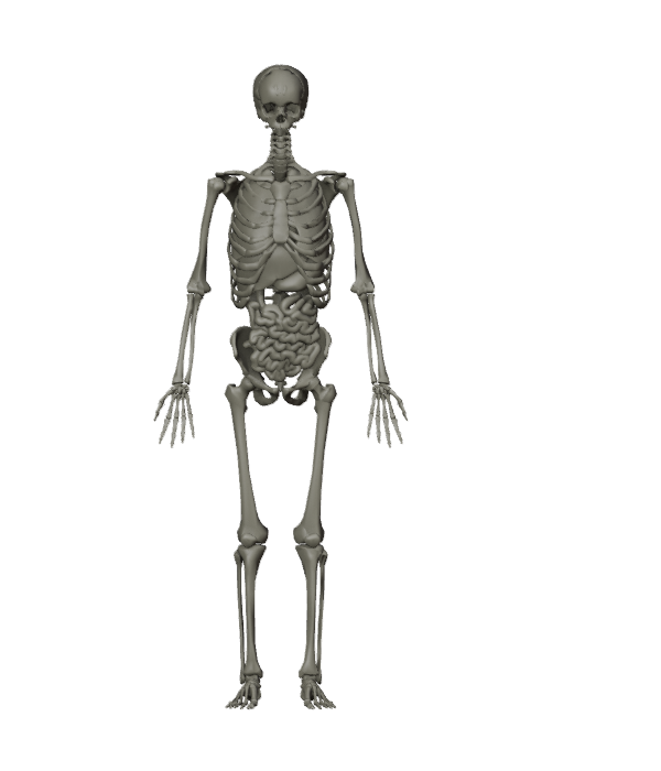
The body contains connected structures and systems. Different imaging methods reveal different kinds of information about them.
Structure and context
Bones provide a framework. Organs and soft tissues sit within and around that framework. A single view cannot show every structure clearly.
What a model can show
This illustration gives a sense of location and relationships. It cannot show what a scan would reveal about your health. Scan coverage depends on the selected service.
The machines
Three ways to look closer.
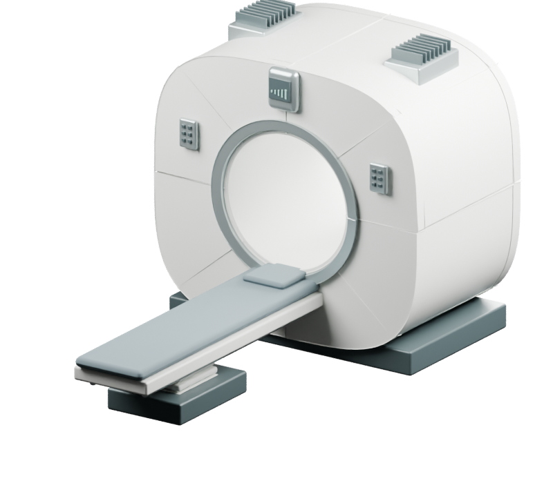
MRI
Magnetic fields and radio signals form images of internal structures.
The magnet creates the main field; radiofrequency coils send or receive signals; gradient coils help locate those signals. MRI does not use ionizing radiation, but safety screening remains essential.
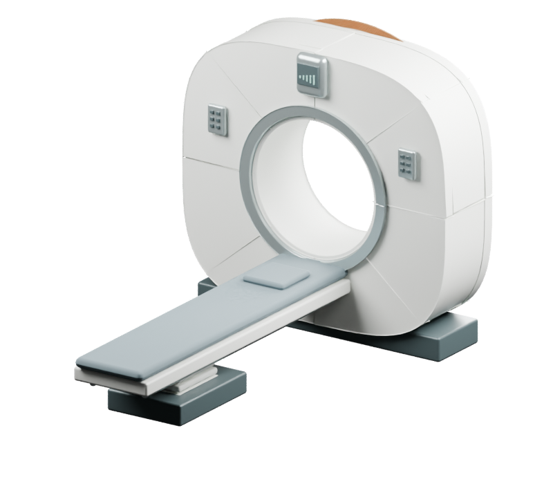
CT
X-ray measurements from many angles are reconstructed into cross-sectional images.
An X-ray source and detectors rotate around the opening while the table positions the body. CT uses ionizing radiation.
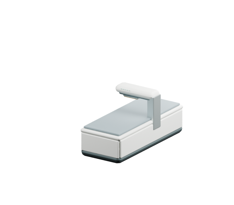
DXA
Two X-ray energies are used to estimate bone mineral density.
A scanning arm contains the source and detector arrangement. DXA uses ionizing radiation and does not produce the same slice-style image stack as MRI or CT.
Models are illustrative. MRI appearance reference: SIGNA Artist Evo; installed model unconfirmed. Internal layouts and animation details in the interactive view are simplified.
The image
One body. Different planes.
These schematic lines show how a viewing plane divides the body. They are orientation diagrams, not scan images.
Axial
Upper and lower portions.
Coronal
Front and back portions.
Sagittal
Left and right portions.
MRI and CT image volumes can be viewed in different planes. The interactive outline is an intersection of modeled surfaces; it does not represent clinical signal intensity.
The body · Interactive atlasExplore beneath the surface.
Keyboard & viewing help
Use Tab to move between controls and Enter or Space to activate them. Use Browse or Search to find a structure without selecting it in the 3D view. Escape closes a detail panel; Tab continues through and out of the explorer.
Prefer a reading view? Choose “Read without 3D” at the beginning of this page.
Choose structures, reveal body systems, or separate the full assembly. This educational atlas shows selected anatomy; scan coverage depends on the selected EverVita protocol.
LOOK CLOSER. GO DEEPER.
A different lens on you
To see beneath the surface, we turn to the science of imaging.
The machines
Meet the machines behind the images.
Choose a machine, take it apart, and explore what each part does.
Keyboard & viewing help
Use Tab to reach Guide me or Explore freely, then Enter or Space to choose. Guide me advances with Next. In Explore freely, use the parts list to select a part without clicking the model.
Prefer a reading view? Choose “Read without 3D” at the beginning of this page.
Different tools. Different information.
Three ways to look closer.
MRI
Magnetic fields + radio signals → images of internal structures.
CT
X-rays from many angles → cross-sectional images.
DXA
Two X-ray energies → an estimate of bone mineral density.
These methods provide different information. The appropriate examination depends on the clinical question.
MRI and CT can produce image volumes viewed in different planes. Move through this model to explore where a cross-section would fall.
Keyboard & viewing help
Use Tab to choose a plane and reach Slice position. Arrow keys move one slice; Home and End move to the first and last slice. Previous and Next provide the same control.
Prefer a reading view? Choose “Read without 3D” at the beginning of this page.
From the machine to the image
Why look in slices?
The anatomy and machines you saw are connected: the same body geometry is sampled in planes, then interpreted into image views by software. This explorer simulates that sampling process.
MRI
Magnetic fields and radio signals make measurements that form image volumes. Those volumes can be viewed in multiple directions.